

High-resolution diffusion tensor imaging in the substantia nigra of de novo Parkinson disease
- PMID: 19129507
- PMCID: PMC2677508
- DOI: 10.1212/01.wnl.0000340982.01727.6e
High-resolution diffusion tensor imaging in the substantia nigra of de novo Parkinson disease
Erratum in
- Neurology. 2009 Jun 9;72(23):2059
Abstract
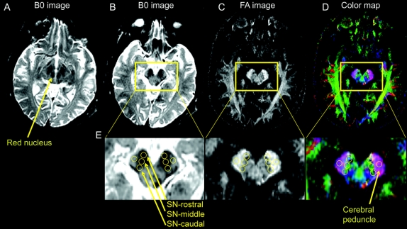
Background: In the midbrain of patients with Parkinson disease (PD), there is a selective loss of dopaminergic neurons in the ventrolateral and caudal substantia nigra (SN). In a mouse model of PD, investigators have administered 1-methyl-4-phenyl-1,2,3,6-tetrahydropyridine (MPTP) and found that measures derived using diffusion tensor imaging (DTI) were correlated with the number of dopamine neurons lost following intoxication.
Methods: Twenty-eight subjects (14 with early stage, untreated PD and 14 age- and gender-matched controls) were studied with a high-resolution DTI protocol at 3 Tesla using an eight-channel phase array coil and parallel imaging to study specific segments of degeneration in the SN. Regions of interest were drawn in the rostral, middle, and caudal SN by two blinded and independent raters.
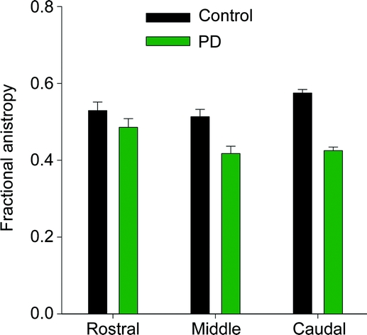
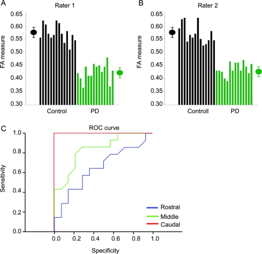
Results: Fractional anisotropy (FA) was reduced in the SN of subjects with PD compared with controls (p < 0.001). Post hoc analysis identified that reduced FA for patients with PD was greater in the caudal compared with the rostral region of interest (p < 0.00001). A receiver operator characteristic analysis in the caudal SN revealed that sensitivity and specificity were 100% for distinguishing patients with PD from healthy subjects. Findings were consistent across both raters.
Conclusions: These findings provide evidence that high resolution diffusion tensor imaging in the substantia nigra distinguishes early stage, de novo patients with Parkinson disease (PD) from healthy individuals on a patient by patient basis and has the potential to serve as a noninvasive early biomarker for PD.
Figures
Comment in
-
A new sensitive imaging biomarker for Parkinson disease?Neurology. 2009 Apr 21;72(16):1374-5. doi: 10.1212/01.wnl.0000343512.36654.41. Epub 2009 Jan 7. Neurology. 2009. PMID: 19129504 No abstract available.
References
-
- Hodaie M, Neimat JS, Lozano AM. The dopaminergic nigrostriatal system and Parkinson’s disease: molecular events in development, disease, and cell death, and new therapeutic strategies. Neurosurgery 2007;60:17–28; discussion 28–30. - PubMed
-
- Braak H, Del Tredici K, Rub U, de Vos RA, Jansen Steur EN, Braak E. Staging of brain pathology related to sporadic Parkinson’s disease. Neurobiol Aging 2003;24:197–211. - PubMed
-
- Fearnley JM, Lees AJ. Ageing and Parkinson’s disease: substantia nigra regional selectivity. Brain 1991;114:2283–2301. - PubMed
-
- Martin WR, Wieler M, Gee M. Midbrain iron content in early Parkinson disease: a potential biomarker of disease status. Neurology 2008;70:1411–1417. - PubMed
Publication types
MeSH terms
Substances
Grants and funding
LinkOut - more resources
Full Text Sources
Other Literature Sources
Medical