. 2009 Apr;80(4):804-12.
doi: 10.1095/biolreprod.108.072603. Epub 2009 Jan 7.

Dynamic nuclear organization of constitutive heterochromatin during fetal male germ cell development in mice

Affiliations

Dynamic nuclear organization of constitutive heterochromatin during fetal male germ cell development in mice

Hirotaka Yoshioka et al. Biol Reprod. 2009 Apr.

Abstract

In mice, male germ cells enter mitotic arrest beginning at 13.5 days postcoitum (dpc), and remain suspended in the G(0)/G(1) cell cycle stage until after birth. During this period, male germ cells undergo extensive epigenetic reprogramming, which is essential for their subsequent function as male gametes. A global reorganization and spatial clustering of constitutive heterochromatin has been implicated in epigenetic plasticity during cellular differentiation. Here, we have studied the dynamics of heterochromatin in fetal (12.5-19.5 dpc) and neonatal (4 days postpartum) male germ cells. We monitored constitutive heterochromatin-specific markers, and observed changes in the association of histone H3 trimethylation of lysine 9 (H3K9me3), binding of heterochromatin protein 1, and patterns of 4',6-diamino-2-phenylindole staining in pericentric regions of chromosomes, along with a coincident loss of chromocenters in fetal prospermatogonia during mitotic arrest. We also observed a transient loss of H3K9me3 associated with major and minor satellite repeat sequences, plus inactivation of histone methyltransferases (Suv39h1 and Suv39h2), and transient activation of histone demethylase (Jmjd2b) in these same cells. These epigenetic changes were correlated with relocation of centromeric regions toward the nuclear periphery in prospermatogonia during mitotic arrest. Taken together, these results show that constitutive heterochromatin undergoes dramatic reorganization during prespermatogenesis. We suggest that these dynamic changes in heterochromatin contribute to normal epigenetic reprogramming of the paternal genome in fetal prospermatogonia suspended in the G(0)/G(1) stage, and that this also represents an epigenomic state that is particularly amenable to reprogramming.


Figures
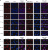
FIG. 1.
FIG. 1.
Dynamic organization of epigenetic heterochromatin states during prespermatogenesis. A) The kinetics of H3K9me3 in fetal and neonatal male germ cells. Male germ cells were isolated from fetuses at 12.5, 13.5, 15.5, 17.5, and 19.5 dpc, and neonates at 4 dpp, and stained with anti-H3K9me3 antibody (red) and DAPI (blue). Arrowheads in the panels of 12.5, 13.5, and 15.5 dpc cells indicate the typical staining patterns of the H3K9me3 as punctate, punctate perinuclear distribution, and diffuse, respectively. B) The kinetics of CBX5 in fetal and neonatal male germ cells. Male germ cells were stained with anti-CBX5 antibody (red) and DAPI (blue). Arrowheads in the panels of 12.5, 13.5, and 15.5 dpc cells indicate the typical staining patterns for CBX5 as punctate, punctate perinuclear distribution, and diffuse, respectively. C and D) Immunofluorescence detection of H3K9me3 (left panels) and CBX5 (right panels) in cumulus cells (C) and in ES cells (D). Bars = 10 μm.
FIG. 2.
FIG. 2.
The proportion of different epigenetic states during male germ cell development. A) The percentage of cells showing positive H3K9me3 staining in a punctate pattern (black bars), a punctate perinuclear distribution (gray bars), or a diffuse distribution (white bars) is shown. B) The percentage of cells showing positive CBX5 staining in a punctate pattern (black bars), a punctate perinuclear distribution (gray bars), or a diffuse distribution (white bars) is shown. At least two independent immunostaining experiments were performed and on ≥ 100 cells each were recorded to calculate the percentages.
FIG. 3.
FIG. 3.
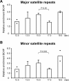
Histone methylation levels at pericentric heterochromatin regions in fetal male germ cells. ChIP analyses were performed on more than 95% purified populations of fetal male germ cells recovered from specific stages prior to and during the period of mitotic arrest using an antibody specific for H3K9me3. Precipitated DNA was subjected to real-time PCR with the use of specific primers for the major satellite repeats (A) and minor satellite repeats (B), and enrichment of immunoprecipitated DNA (bound fractions [BO]) relative to input (IP) DNA was calculated in each case. Data represent mean (± SEM) values of two independent ChIP experiments for 12.5 dpc and 19.5 dpc, and more than three for 13.5 dpc, 15.5 dpc, and 17.5 dpc. Two dots show the extent of variation when only two measurements were made. Statistical analyses were performed using the Student t-test. A value of P < 0.05 was considered statistically significant.
FIG. 4.
FIG. 4.
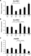
Quantitative expression of two histone methyltransferase genes and a histone demethylase gene during fetal and neonatal male germ cell development. Relative expression of two different histone methyltransferase genes, Suv39h1 (A) and Suv39h2 (B), and a histone demethylase, Jmjd2b (C), in fetal and neonatal male germ cells are shown. In each case, expression of each gene was measured in endogenous male germ cells (recovered at 12.5, 13.5, 15.5, 17.5, and 19.5 dpc, and 4 dpp, and purified to 100%). The relative expression of each target mRNA was calculated from the target CT values and Actb mRNA CT values, using the standard curve method. The relative amount of each target mRNA at 12.5 dpc was arbitrarily set at 1, and all other transcript levels were compared to these values. Error bars indicate SEM. Statistical analyses were performed using the Student t-test. A value of P < 0.05 was considered statistically significant.
FIG. 5.
FIG. 5.
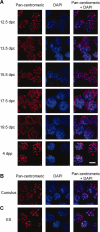
Changes in nuclear architecture during prespermatogenesis. A) Dynamic reorganization of pericentric heterochromatin domains during fetal and neonatal male germ cell development. Male germ cells were isolated from fetuses at 12.5, 13.5, 15.5, 17.5, and 19.5 dpc, and neonates at 4 dpp, and hybridized with a Cy3-labeled pancentromeric probe (red) to visualize the nuclear localization of centromeric regions. DNA was stained with DAPI (blue). B and C) Localization of centromeric regions in cumulus cells (B) and in ES cells (C). Bar = 10 μm.

References

    1. Ohinata Y, Payer B, O'Carroll D, Ancelin K, Ono Y, Sano M, Barton SC, Obukhanych T, Nussenzweig M, Tarakhovsky A, Saitou M, Surani MA.Blimp1 is a critical determinant of the germ cell lineage in mice. Nature 2005; 436: 207–213. - PubMed
    1. McCarrey JR.Development of the germ cell. Desjardins C, Ewing LL.Cell and Molecular Biology of the Testis New York:Oxford University Press;1993: 58–89.
    1. McLaren A.Primordial germ cells in the mouse. Dev Biol 2003; 262: 1–15. - PubMed
    1. Koopman P, Münsterberg A, Capel B, Vivian N, Lovell-Badge R.Expression of a candidate sex-determining gene during mouse testis differentiation. Nature 1990; 348: 450–452. - PubMed
    1. Wilhelm D, Koopman P.The makings of maleness: towards an integrated view of male sexual development. Nat Rev Genet 2006; 7: 620–631. - PubMed

Publication types

MeSH terms