

Tissue microarrays of human tumor xenografts: characterization of proteins involved in migration and angiogenesis for applications in the development of targeted anticancer agents
- PMID: 19129557
- PMCID: PMC3662302
Tissue microarrays of human tumor xenografts: characterization of proteins involved in migration and angiogenesis for applications in the development of targeted anticancer agents
Abstract
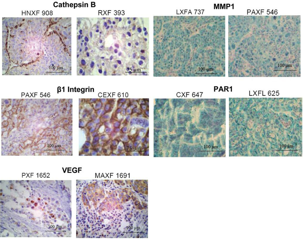
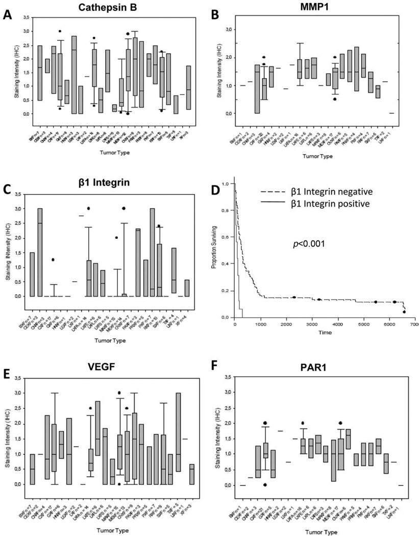
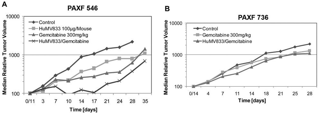
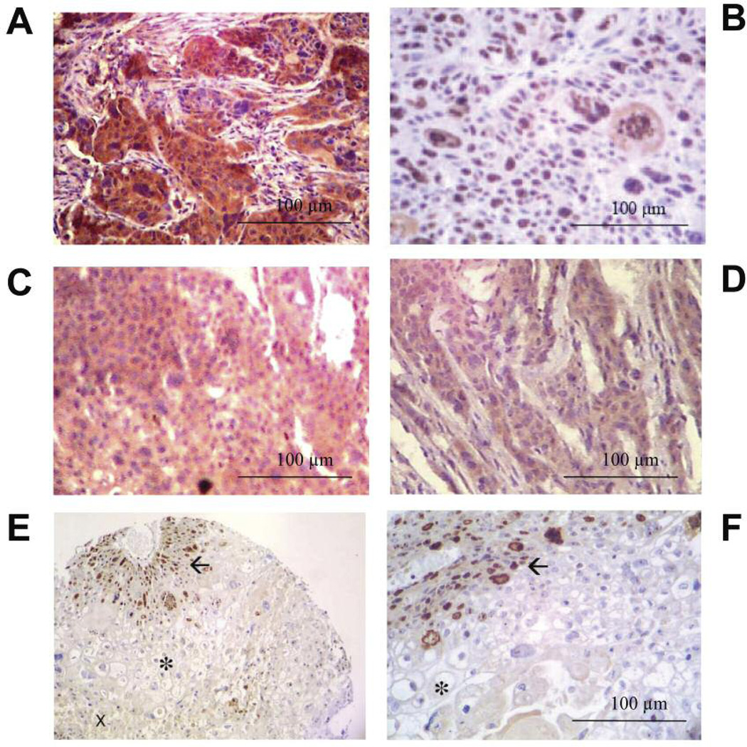
As new target-directed anticancer agents emerge, preclinical efficacy studies need to integrate target-driven model systems. This approach to drug development requires rapid and reliable characterization of the new targets in established tumor models, such as xenografts and cell lines. Here, we have applied tissue microarray technology to patient-derived, re-growable human tumor xenografts. We have profiled the expression of five proteins involved in cell migration and/or angiogenesis: vascular endothelial growth factor (VEGF), matrix metalloproteinase 1 (MMP1), protease activated receptor (PAR1), cathepsin B, and beta1 integrin in a panel of over 150 tumors and compared their expression levels to available patient outcome data. For each protein, several target overexpressing xenografts were identified. They represent a subset of tumor models prone to respond to specific inhibitors and are available for future preclinical efficacy trials. In a "proof of concept" experiment, we have employed tissue microarrays to select in vivo models for therapy and for the analysis of molecular changes occurring after treatment with the anti-VEGF antibody HuMV833 and gemcitabine. Whereas the less angiogenic pancreatic cancer PAXF736 model proved to be resistant, the highly vascularized PAXF546 xenograft responded to therapy. Parallel analysis of arrayed biopsies from the different treatment groups revealed a down-regulation of Ki-67 and VEGF, an altered tissue morphology, and a decreased vessel density. Our results demonstrate the multiple advantages of xenograft tissue microarrays for preclinical drug development.
Figures
Similar articles
-
Effect of the vascular endothelial growth factor receptor-2 antibody DC101 plus gemcitabine on growth, metastasis and angiogenesis of human pancreatic cancer growing orthotopically in nude mice.Int J Cancer. 2002 Nov 10;102(2):101-8. doi: 10.1002/ijc.10681. Int J Cancer. 2002. PMID: 12385004
-
Specific targeting of tumor endothelial cells by a shiga-like toxin-vascular endothelial growth factor fusion protein as a novel treatment strategy for pancreatic cancer.Neoplasia. 2010 Oct;12(10):797-806. doi: 10.1593/neo.10418. Neoplasia. 2010. PMID: 20927318 Free PMC article.
-
Antiangiogenic versus cytotoxic therapeutic approaches to human pancreas cancer: an experimental study with a vascular endothelial growth factor receptor-2 tyrosine kinase inhibitor and gemcitabine.Eur J Pharmacol. 2004 Sep 13;498(1-3):9-18. doi: 10.1016/j.ejphar.2004.07.062. Eur J Pharmacol. 2004. PMID: 15363970
-
Tumor angiogenesis and accessibility: role of vascular endothelial growth factor.Semin Oncol. 2002 Dec;29(6 Suppl 16):3-9. doi: 10.1053/sonc.2002.37265. Semin Oncol. 2002. PMID: 12516032 Review.
-
[Molecular-based treatment concepts in advanced pancreatic cancer].Dtsch Med Wochenschr. 2007 Apr 13;132(15):818-22. doi: 10.1055/s-2007-973627. Dtsch Med Wochenschr. 2007. PMID: 17427093 Review. German. No abstract available.
Cited by
-
Llama-derived single variable domains (nanobodies) directed against chemokine receptor CXCR7 reduce head and neck cancer cell growth in vivo.J Biol Chem. 2013 Oct 11;288(41):29562-72. doi: 10.1074/jbc.M113.498436. Epub 2013 Aug 26. J Biol Chem. 2013. PMID: 23979133 Free PMC article.
-
Activation of Epidermal Growth Factor Receptor/p38/Hypoxia-inducible Factor-1α Is Pivotal for Angiogenesis and Tumorigenesis of Malignantly Transformed Cells Induced by Hexavalent Chromium.J Biol Chem. 2016 Jul 29;291(31):16271-81. doi: 10.1074/jbc.M116.715797. Epub 2016 May 25. J Biol Chem. 2016. Retraction in: J Biol Chem. 2019 Oct 18;294(42):15558. doi: 10.1074/jbc.RX119.011168. PMID: 27226640 Free PMC article. Retracted.
-
Ex Vivo Testing of Patient-Derived Xenografts Mirrors the Clinical Outcome of Patients with Pancreatic Ductal Adenocarcinoma.Clin Cancer Res. 2016 Dec 15;22(24):6021-6030. doi: 10.1158/1078-0432.CCR-15-2936. Epub 2016 Jun 3. Clin Cancer Res. 2016. PMID: 27259561 Free PMC article.
-
Patient-derived tumour xenografts as models for oncology drug development.Nat Rev Clin Oncol. 2012 Apr 17;9(6):338-50. doi: 10.1038/nrclinonc.2012.61. Nat Rev Clin Oncol. 2012. PMID: 22508028 Free PMC article. Review.
-
Targeted therapy for head and neck cancer: signaling pathways and clinical studies.Signal Transduct Target Ther. 2023 Jan 16;8(1):31. doi: 10.1038/s41392-022-01297-0. Signal Transduct Target Ther. 2023. PMID: 36646686 Free PMC article. Review.
References
-
- Burger AM. Highlights in experimental therapeutics. Cancer Lett. 2007;245:11–21. - PubMed
-
- Burger AM, Fiebig HH. Preclinical screening for new anticancer agents. In: Figg WD, McLeod HL, editors. Handbook of Anticancer Pharmacokinetics and Pharmacodynamics. Totowa, NJ: Humana Press Inc; 2004. pp. 29–44.
-
- Kononen J, Bubendorf L, Kallioniemi A, Bärlund M, Scraml P, Leighton S, et al. Tissue microarrays for high-throughput molecular profiling of tumor specimens. Nat Med. 1998;4:844–847. - PubMed
-
- Fiebig HH, Maier A, Burger AM. Clonogenic assay with established human tumor xenografts: correlation of in vitro to in vivo activity as a basis for anticancer drug discovery. Eur J Cancer. 2004;40:802–820. - PubMed
Publication types
MeSH terms
Substances
Grants and funding
LinkOut - more resources
Full Text Sources
Medical
Research Materials