. 2009 Jan 22;457(7228):446-50.
doi: 10.1038/nature07637. Epub 2009 Jan 7.

Intersubunit coordination in a homomeric ring ATPase

Affiliations

Intersubunit coordination in a homomeric ring ATPase

Jeffrey R Moffitt et al. Nature. .

Abstract

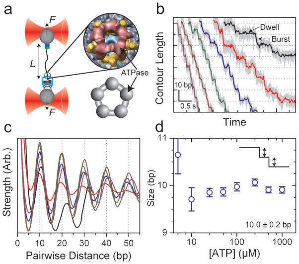
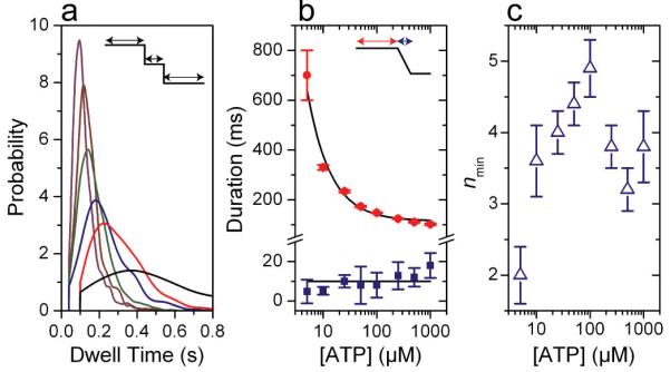
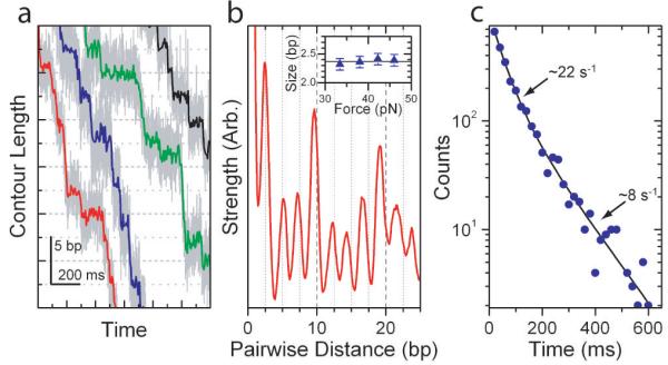
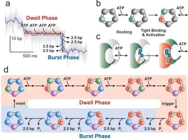
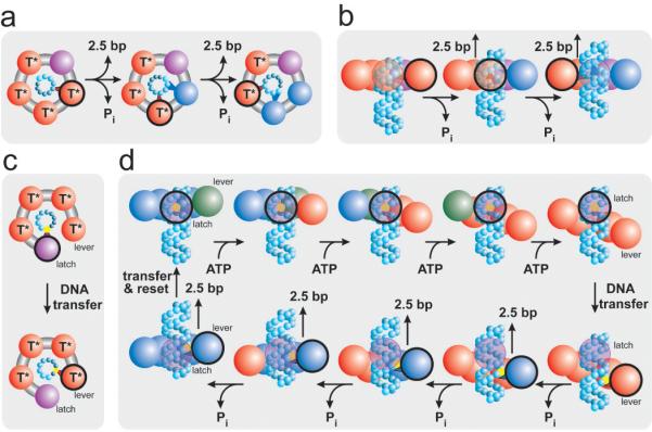
Homomeric ring ATPases perform many vital and varied tasks in the cell, ranging from chromosome segregation to protein degradation. Here we report the direct observation of the intersubunit coordination and step size of such a ring ATPase, the double-stranded-DNA packaging motor in the bacteriophage phi29. Using high-resolution optical tweezers, we find that packaging occurs in increments of 10 base pairs (bp). Statistical analysis of the preceding dwell times reveals that multiple ATPs bind during each dwell, and application of high force reveals that these 10-bp increments are composed of four 2.5-bp steps. These results indicate that the hydrolysis cycles of the individual subunits are highly coordinated by means of a mechanism novel for ring ATPases. Furthermore, a step size that is a non-integer number of base pairs demands new models for motor-DNA interactions.


Figures

Figure 1
Figure 1. Bacteriophage φ29 Packages DNA in Bursts of 10 bp
(a) A single packaging bacteriophage prohead-motor complex and its dsDNA substrate are tethered between two beads each held in an optical trap. Inset: cryo-EM reconstruction of the full motor complex (Courtesy of M. Morais), ATPase in red, pRNA in yellow, connector in cyan, and capsid in gray with a top view cartoon of the ATPase ring alone (below, gray). (b) Representative packaging traces collected under low external load, ∼8 pN, and different [ATP]: 250 μM, 100 μM, 50 μM, 25 μM, 10 μM, and 5 μM in purple, brown, green, blue, red, and black, respectively, all boxcar-filtered and decimated to 50 Hz. Data at 1.25 kHz are plotted in light gray. (c) Average pairwise distributions of packaging traces selected for low noise levels (50% of all packaging data; see Supplementary Figures 2 and 3). Color scheme as in (b). (d) The average size of the packaging burst versus [ATP] determined from the periodicity in (c). Error bars are the error in the slope from a linear fit to the peak position. Data collected at 500 μM and 1 mM [ATP] are not shown in (b) and (c) for clarity.
Figure 2
Figure 2. Dwells Before 10-bp Bursts Contain Multiple Kinetic Events
(a) Probability distributions for the dwell times preceding a 10-bp burst under low external load, ∼8 pN, and different [ATP]: color scheme as in Figure 1. Distributions were estimated using kernel density estimation with a Gaussian kernel and the optimum bandwidth and are truncated at the lowest detectable dwell time. Supplementary Figure 2 contains the number of observed bursts for each [ATP]. Distributions for 500 μM and 1 mM [ATP] are not shown for clarity. (b) The mean dwell time before the 10-bp bursts (red circles) for all [ATP] with an inverse hyperbolic fit (black) and the mean duration of all bursts (blue squares, average denoted by black line). (c) The minimum number of rate-limiting kinetic events during the dwell before the 10-bp bursts, nmin, for all [ATP]. Error bars are the standard error.
Figure 3
Figure 3. The 10-bp Bursts are Composed of Four 2.5-bp Steps
(a) Representative packaging traces collected with external loads of ∼40 pN and 250 μM [ATP]. Data in light gray are plotted at 1.25 kHz while data in color are boxcar-filtered and decimated to 100 Hz. (b) Average pairwise distribution of packaging traces selected for low noise levels (50% of all packaging data; see Supplementary Figures 2 and 3). Inset: Force dependence of the observed spatial periodicity. The solid line is the mean for all forces 2.4±0.1 bp (s.e.m.). (c) Dwell time histogram for the 2.5-bp steps observed under the packaging conditions seen in (a) plotted in blue circles with a bi-exponential fit in black (N=2,662).
Figure 4
Figure 4. Inter-Subunit Coordination in the Ring-ATPase of φ29
(a) Schematic diagram of the two-phase mechanochemical cycle of φ29 overlaid on a sample packaging trace. (b) Detailed kinetics of ATP binding. Binding occurs in two steps, ATP docking (green, T) followed by tight-binding (red, T*). (c) Schematic diagram of the communication between subunits during ATP binding. Upon tight-binding of an ATP, the binding pocket of the next subunit, formerly inactive (gray), is activated for docking (green). (d) Schematic depiction of the full mechanochemical cycle of φ29. During the burst phase, ADP may remain on the ring (blue) to be released in the dwell phase. One subunit must be distinct from the others (purple) in order to break the symmetry of the motor and generate only 4 steps per cycle. The identity of this subunit may change each cycle.
Figure 5
Figure 5. Packaging Models that Produce a Non-Integer Step Size
(a) Depiction of a translocation model in which all subunits eventually contact the DNA (cyan spheres.) The contacting subunit is outlined in black. (b) In such a model the size of internal conformational changes set the step size (side view.) (c) Depiction of a translocation model in which only two subunits contact the DNA (black outline.) (d) In such a model, one subunit maintains contact with the DNA (the latch) while the loading of each ATP introduces relative subunit-subunit rotations which distort the ring. This distortion extends one subunit (the lever) along the DNA by ∼10-bp. The DNA contact point is then transferred from the latch to the lever, and the release of hydrolysis products relaxes the ring, retracting the lever and the DNA. The DNA contact is then transferred back to the latch, the ring resets, and the cycle begins again. Because there are four subunits, the ring is retracted in four steps, dividing a 10-bp step into four ∼2.5-bp substeps. The subunit color scheme is the same as in Figure 4.

Comment in

References

    1. Latterich M, Patel S. The AAA team: related ATPases with diverse functions. Trends in Cell Biology. 1998;8:65. - PubMed
    1. Ogura T, Wilkinson AJ. AAA+ superfamily ATPases: common structure--diverse function. Genes to Cells. 2001;6:575–597. - PubMed
    1. Iyer LM, Leipe DD, Koonin EV, Aravind L. Evolutionary history and higher order classification of AAA+ ATPases. J Struct Biol. 2004;146:11–31. - PubMed
    1. Kainov DE, Tuma R, Mancini EJ. Hexameric molecular motors: P4 packaging ATPase unravels the mechanism. Cell Mol Life Sci. 2006;63:1095–105. - PMC - PubMed
    1. Erzberger JP, Berger JM. Evolutionary relationships and structural mechanisms of AAA+ proteins. Annu Rev Biophys Biomol Struct. 2006;35:93–114. - PubMed

Publication types