A molecular imaging paradigm to rapidly profile response to angiogenesis-directed therapy in small animals
- PMID: 19130143
- PMCID: PMC2677126
- DOI: 10.1007/s11307-008-0193-9
A molecular imaging paradigm to rapidly profile response to angiogenesis-directed therapy in small animals
Abstract
Purpose: The development of novel angiogenesis-directed therapeutics is hampered by the lack of non-invasive imaging metrics capable of assessing treatment response. We report the development and validation of a novel molecular imaging paradigm to rapidly assess response to angiogenesis-directed therapeutics in preclinical animal models.
Procedures: A monoclonal antibody-based optical imaging probe targeting vascular endothelial growth factor receptor-2 (VEGFR2) expression was synthesized and evaluated in vitro and in vivo via multispectral fluorescence imaging.
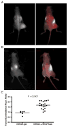
Results: The optical imaging agent demonstrated specificity for the target receptor in cultured endothelial cells and in vivo. The agent exhibited significant accumulation within 4T1 xenograft tumors. Mice bearing 4T1 xenografts and treated with sunitinib exhibited both tumor growth arrest and decreased accumulation of NIR800-alphaVEGFR2ab compared to untreated cohorts (p = 0.0021).
Conclusions: Molecular imaging of VEGFR2 expression is a promising non-invasive biomarker for assessing angiogenesis and evaluating the efficacy of angiogenesis-directed therapies.
Figures


Similar articles
-
Inhibition of Endothelial SCUBE2 (Signal Peptide-CUB-EGF Domain-Containing Protein 2), a Novel VEGFR2 (Vascular Endothelial Growth Factor Receptor 2) Coreceptor, Suppresses Tumor Angiogenesis.Arterioscler Thromb Vasc Biol. 2018 May;38(5):1202-1215. doi: 10.1161/ATVBAHA.117.310506. Epub 2018 Mar 15. Arterioscler Thromb Vasc Biol. 2018. PMID: 29545238
-
SKLB610: a novel potential inhibitor of vascular endothelial growth factor receptor tyrosine kinases inhibits angiogenesis and tumor growth in vivo.Cell Physiol Biochem. 2011;27(5):565-74. doi: 10.1159/000329978. Epub 2011 Jun 15. Cell Physiol Biochem. 2011. PMID: 21691074
-
YM-359445, an orally bioavailable vascular endothelial growth factor receptor-2 tyrosine kinase inhibitor, has highly potent antitumor activity against established tumors.Clin Cancer Res. 2006 Mar 1;12(5):1630-8. doi: 10.1158/1078-0432.CCR-05-2028. Clin Cancer Res. 2006. PMID: 16533791
-
BR55: a lipopeptide-based VEGFR2-targeted ultrasound contrast agent for molecular imaging of angiogenesis.Invest Radiol. 2010 Feb;45(2):89-95. doi: 10.1097/RLI.0b013e3181c5927c. Invest Radiol. 2010. PMID: 20027118
-
Molecular imaging of vessels in mouse models of disease.Eur J Radiol. 2009 May;70(2):305-11. doi: 10.1016/j.ejrad.2009.01.053. Eur J Radiol. 2009. PMID: 19304428 Free PMC article. Review.
Cited by
-
Imaging key biomarkers of tumor angiogenesis.Theranostics. 2012;2(5):502-15. doi: 10.7150/thno.3623. Epub 2012 May 17. Theranostics. 2012. PMID: 22737188 Free PMC article.
-
Molecular imaging metrics to evaluate response to preclinical therapeutic regimens.Front Biosci (Landmark Ed). 2011 Jan 1;16(2):393-410. doi: 10.2741/3694. Front Biosci (Landmark Ed). 2011. PMID: 21196177 Free PMC article. Review.
-
Synthesis and evaluation of aryl boronic acids as fluorescent artificial receptors for biological carbohydrates.Bioorg Chem. 2012 Feb;40(1):137-142. doi: 10.1016/j.bioorg.2011.11.003. Epub 2011 Nov 25. Bioorg Chem. 2012. PMID: 22177855 Free PMC article.
-
In vivo longitudinal imaging of RNA interference-induced endocrine therapy resistance in breast cancer.J Biophotonics. 2020 Jan;13(1):e201900180. doi: 10.1002/jbio.201900180. Epub 2019 Oct 9. J Biophotonics. 2020. PMID: 31595691 Free PMC article.
-
Multispectral fluorescence imaging to assess pH in biological specimens.J Biomed Opt. 2011 Jan-Feb;16(1):016007. doi: 10.1117/1.3533264. J Biomed Opt. 2011. PMID: 21280913 Free PMC article.
References
-
- Folkman J. Angiogenesis in cancer, vascular, rheumatoid and other disease. Nat Med (USA) 1995;1:27–31. - PubMed
-
- Folkman J, Watson K, Ingber D, Hanahan D. Induction of angiogenesis during the transition from hyperplasia to neoplasia. Nature (USA) 1989;339:58–61. - PubMed
-
- Folkman J. Tumor angiogenesis: therapeutic implications. N Engl J Med (USA) 1971;285:1182–1186. - PubMed
-
- Ferrara N, Henzel WJ. Pituitary follicular cells secrete a novel heparin-binding growth factor specific for vascular endothelial cells. Biochem Biophys Res Commun (USA) 1989;161:851–858. - PubMed
Publication types
MeSH terms
Substances
Grants and funding
LinkOut - more resources
Full Text Sources
Other Literature Sources
Medical

