. 2009 Mar;45(3):284-92.
doi: 10.1016/j.ceca.2008.11.006. Epub 2009 Jan 7.

Intracellular calcium signals regulate growth of hepatic stellate cells via specific effects on cell cycle progression

Affiliations

Intracellular calcium signals regulate growth of hepatic stellate cells via specific effects on cell cycle progression

Elwy M Soliman et al. Cell Calcium. 2009 Mar.

Abstract

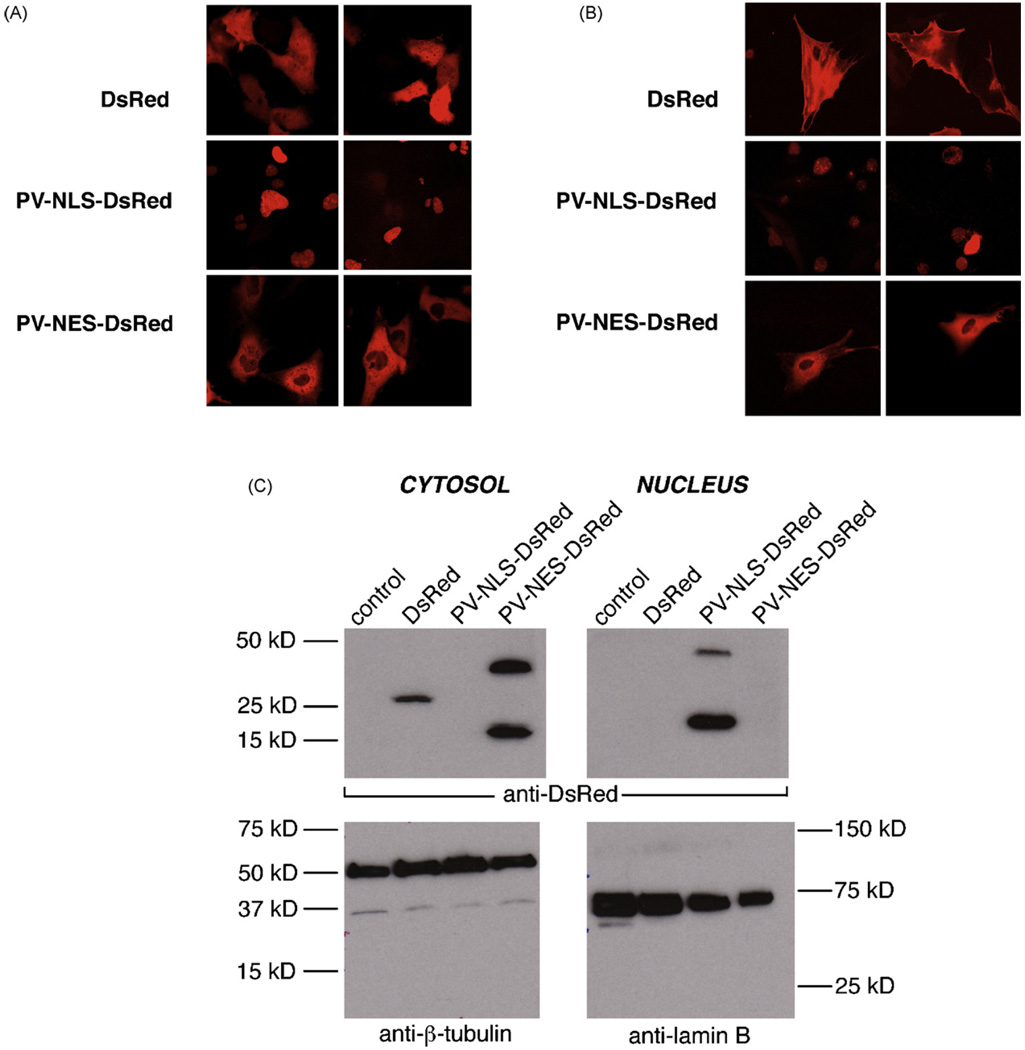
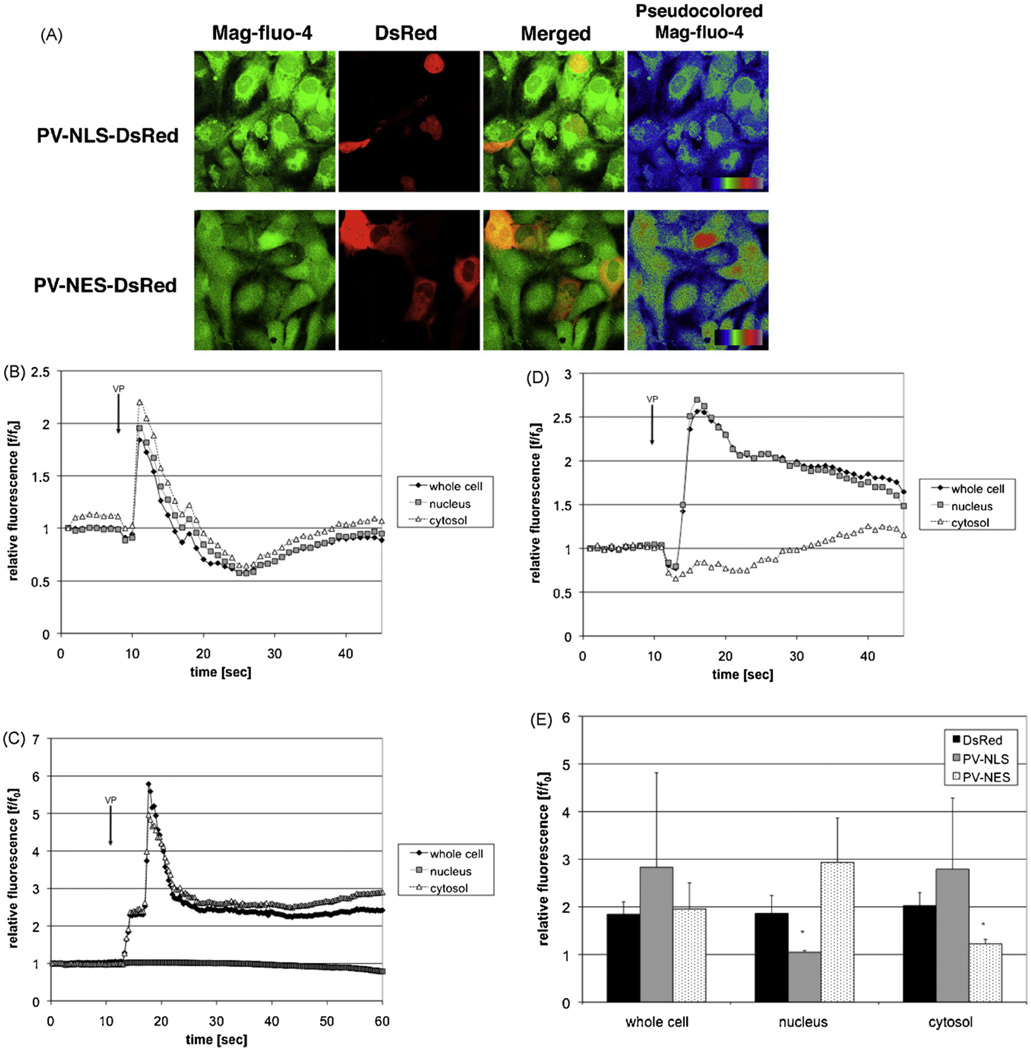
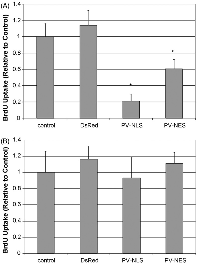
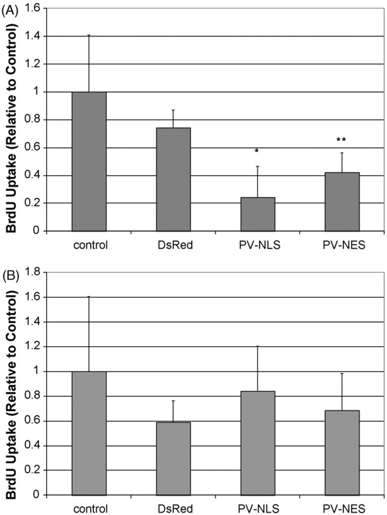
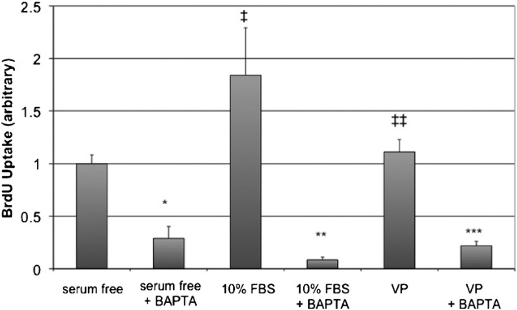
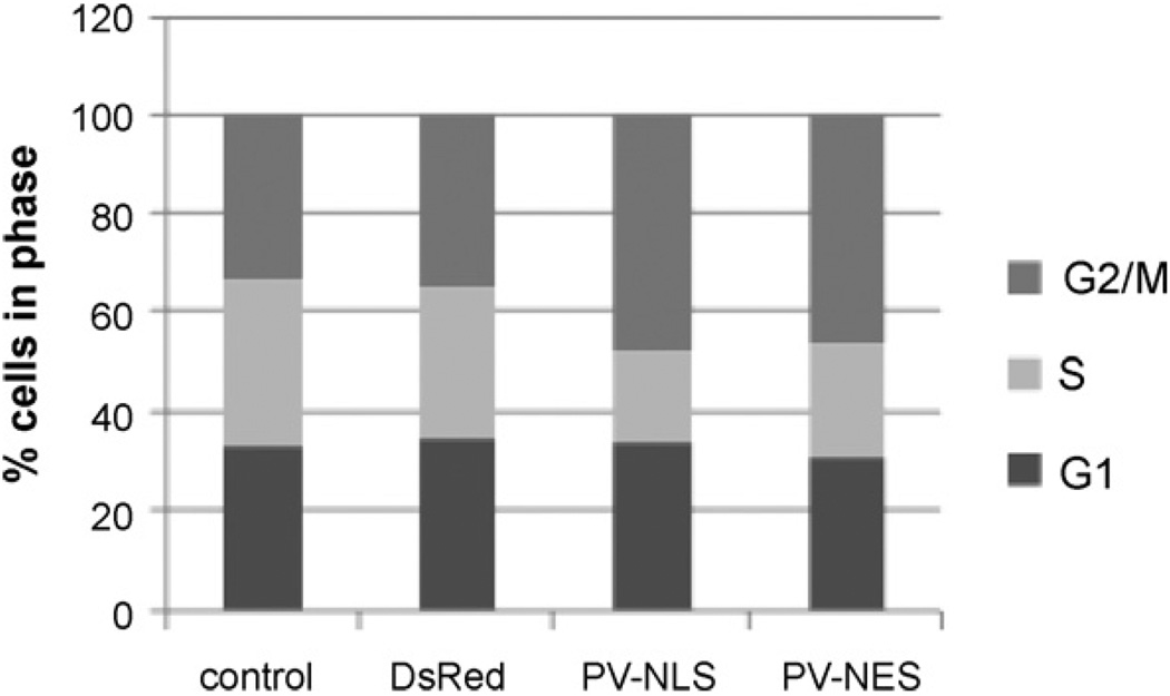
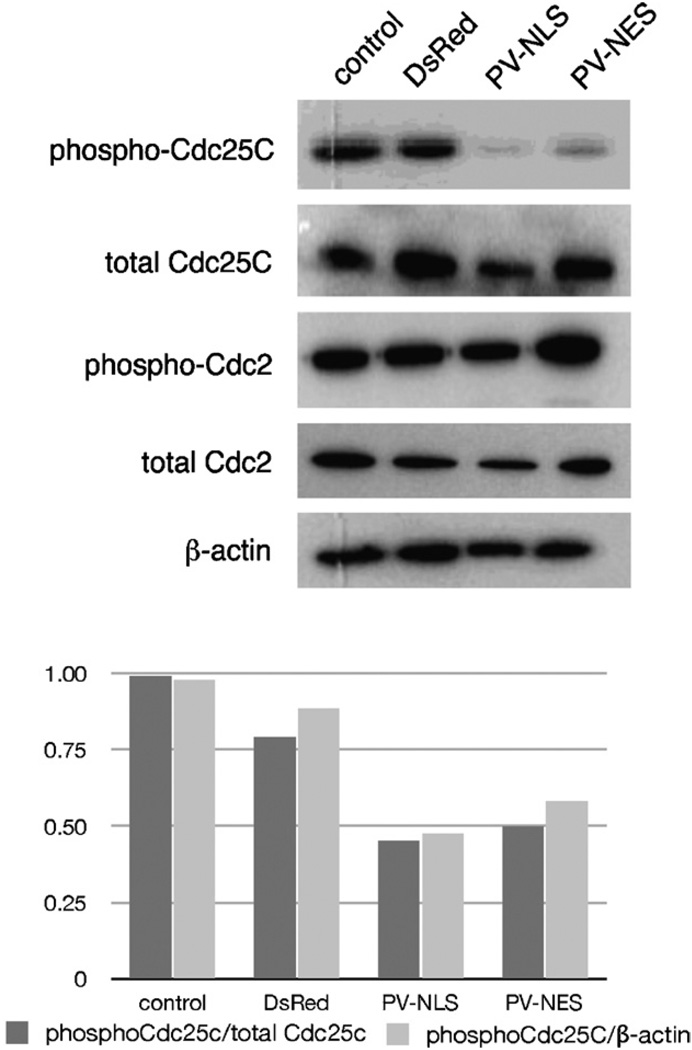
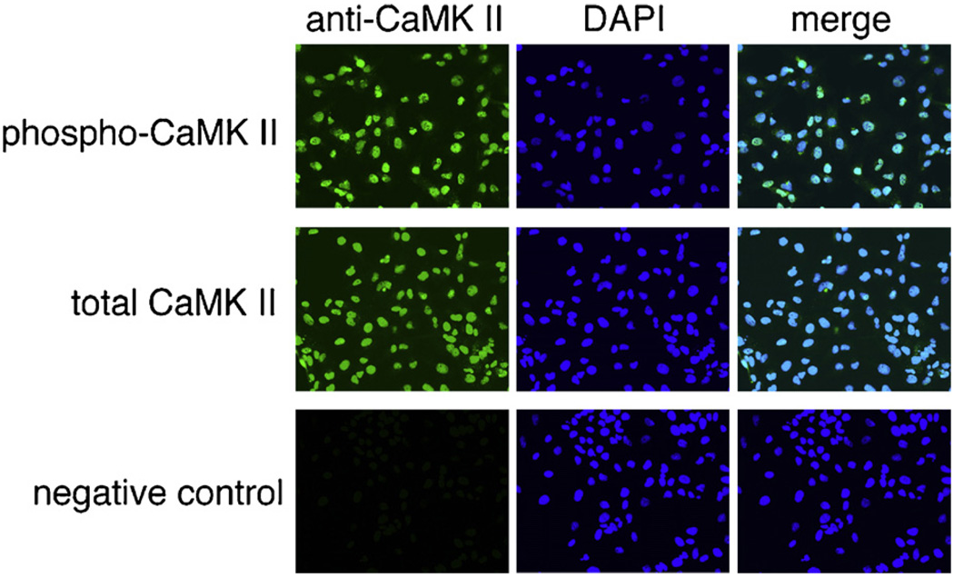
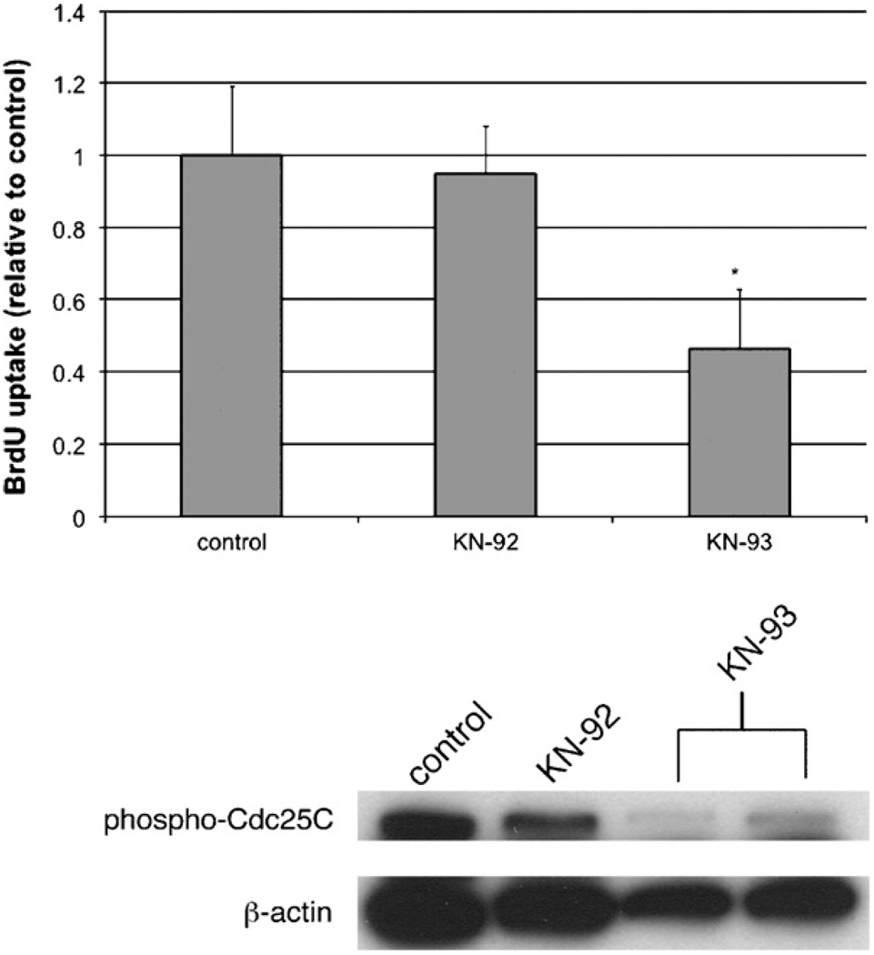
Hepatic stellate cells (HSC) are important mediators of liver fibrosis. Hormones linked to downstream intracellular Ca(2+) signals upregulate HSC proliferation, but the mechanisms by which this occurs are unknown. Nuclear and cytosolic Ca(2+) signals may have distinct effects on cell proliferation, so we expressed plasmid and adenoviral constructs containing the Ca(2+) chelator parvalbumin (PV) linked to either a nuclear localization sequence (NLS) or a nuclear export sequence (NES) to block Ca(2+) signals in distinct compartments within LX-2 immortalized human HSC and primary rat HSC. PV-NLS and PV-NES constructs each targeted to the appropriate intracellular compartment and blocked Ca(2+) signals only within that compartment. PV-NLS and PV-NES constructs inhibited HSC growth. Furthermore, blockade of nuclear or cytosolic Ca(2+) signals arrested growth at the G2/mitosis (G2/M) cell-cycle interface and prevented the onset of mitosis. Blockade of nuclear or cytosolic Ca(2+) signals downregulated phosphorylation of the G2/M checkpoint phosphatase Cdc25C. Inhibition of calmodulin kinase II (CaMK II) had identical effects on LX-2 growth and Cdc25C phosphorylation. We propose that nuclear and cytosolic Ca(2+) are critical signals that regulate HSC growth at the G2/M checkpoint via CaMK II-mediated regulation of Cdc25C phosphorylation. These data provide a new logical target for pharmacological therapy directed against progression of liver fibrosis.


Conflict of interest statement

Conflict of interest statement

The authors have no conflicts of interest that could inappropriately influence the work.

Figures

Fig. 1
Fig. 1
PV constructs can be targeted to subcellular regions within LX-2 cells and primary myofibroblastic HSC. (A) Expression in LX-2 cells. LX-2 cells were transfected with DsRed, PV-NLS-DsRed, or PV-NES-DsRed, and fluorescence distribution was determined by confocal microscopy. Two representative images are seen for each condition. As seen in the upper figure, DsRed fluorescence is seen throughout the cell. On the other hand, as seen in the middle figure, PV-NLS-DsRed fluorescence is limited to the nucleus, and PV-NES-DsRed fluorescence is limited to the extra-nuclear cytoplasm. (B) Expression in primary rat HSC. HSC were infected with DsRed, PV-NLS-DsRed, or PV-NES-DsRed, and fluorescence distribution was determined as described above. All cells are shown at 630× magnification. (C) Verification of PV construct targeting in transfected LX-2 cells. LX-2 cells were transfected as described above then saponified. Crude protein extracts were separated into nuclear and cytosolic fractions. Expression of DsRed (predicted MW 30 kD with non-specific band at 20 kD reported by manufacturer), β-tubulin (cytosolic marker, 55 kD), and lamin B (nuclear marker, 68 kD) were determined by immunoblot. DsRed and PV-NES-DsRed were detected only in cytosolic fractions, and PV-NLS-DsRed was detected only in nuclear fractions, demonstrating the specificity of targeting of these constructs. The predicted MW of PV-DsRed constructs is 42 kD, based on a MWof 12 kD for PV and 30 kD for DsRed.
Fig. 2
Fig. 2
Targeted PV constructs block Ca2+ signals within distinct subcellular regions within LX-2 cells. (A) PV constructs do not alter stored Ca2+. LX-2 cells were transfected with PV-NLS-DsRed or PV-NES-DsRed and loaded with the low-affinity Ca2+ fluorophoreMag-fluo-4, which is an indicator of stored Ca2+. As can be seen by the representative confocal images, no change in Mag-fluo-4 fluorescence can be seen in transfected cells. (B–D) Representative tracings of confocal video microscopy experiments. LX-2 cells were transfected with DsRed (B), PV-NLS-DsRed (C), or PV-NES-DsRed (D), and Ca2+ signals were assessed using confocal video microscopy. Representative tracings showing Ca2+ changes over time after perifusion with VP (2 µM) [8] are shown. Simultaneous Ca2+ increases of similar magnitude were observed in cells transfected with DsRed alone. However, in cells transfected with PV-NLS-DsRed, Ca2+ increases were limited to the extra-nuclear cytoplasm. In contrast, cells transfected with PV-NES-DsRed had Ca2+ increases limited to the nucleus. E. Composite of confocal video microscopy experiments. Peak fluorescence/baseline fluorescence ratios (f/f0) were determined for each condition. PV-NLS-DsRed-expressing cells exhibited no increase in Ca2+ within nuclei, and PV-NES-DsRed-expressing cells exhibited no increase in Ca2+ within the cytosol (n = 4 separate perifusions; *p < 0.05 vs. DsRed).
Fig. 3
Fig. 3
Ca2+ chelation within nuclei or extra-nuclear cytoplasm inhibits LX-2 cell proliferation. (A) Proliferation in the presence of serum. LX-2 cells were either untransfected or transfected with DsRed, PV-NLS-DsRed, or PV-NES DsRed, and proliferation was determined by BrdU uptake. Relative to control, proliferation of cells transfected with either PV-NLS-DsRed or PV-NES-DsRed was diminished (*p < 10−5; n = 10 for each condition). (B) Proliferation in the absence of serum. LX-2 cells were transfected identically to those in (A); however, cells were grown in serum-free media. As seen, no change in proliferation was noted.
Fig. 4
Fig. 4
Ca2+ chelation within nuclei or extra-nuclear cytoplasm inhibits proliferation of activated primary rat HSC. (A) Proliferation in the presence of serum. Primary rat HSC were either uninfected or infected with adenoviral vectors containing DsRed, PV-NLS-DsRed, or PV-NES DsRed, and proliferation was determined by BrdU uptake. Relative to control, proliferation of cells transfected with either PV-NLS-DsRed or PV-NES-DsRed was diminished (*p < 0.01; **p < 0.05; n = 5 for each condition). (B) Proliferation in the absence of serum. Primary rat HSC were infected identically to those in (A); however, cells were grown in serum-free media. As seen, no change in proliferation was noted.
Fig. 5
Fig. 5
The Ca2+ chelator BAPTA/AM inhibits LX-2 proliferation. The effect of the cell-permeant Ca2+ chelator BAPTA/AM on cell proliferation was determined in cells under serum-free, serum-treated, and VP-treated conditions. Serum significantly increased LX-2 growth (p < 0.02 vs. control), whereas, VP had no effect (‡‡ p = NS). BAPTA/AM significantly blocked LX-2 growth in all conditions (*p < 10−5 vs. serum-free; **p = 0.001 vs. 10% FBS; ***p < 10−5 vs. VP; n = 5 for all conditions).
Fig. 6
Fig. 6
Ca2+ chelation within nuclei or extra-nuclear cytoplasm arrests HSC at the G2 phase of the cell cycle. LX-2 cells were either untransfected or transfected with DsRed, PV-NLS-DsRed, or PV-NES-DsRed, and cell cycle was determined by FACS analysis. Relative to control or DsRed-transfected cells, the fraction of cells in G2/M phase was increased from 33–35% to 46–48%, with a consequent decrease in cell fraction in S phase from 30–33% to 18–23%. No change in fraction of cells in G1 phase was noted, as the fraction in all cells was 31–35%.
Fig. 7
Fig. 7
Ca2+ chelation within nuclei or extra-nuclear cytoplasm inhibits phosphorylation of the cyclin phosphatase Cdc25C. (A) Immunoblot. LX-2 cells were transfected identically to those in Fig. 5, and expression of phosphorylated and total cyclins was determined by immunoblot. A marked downregulation of phospho-Cdc25C is noted in cells transfected with PV-NLS-DsRed or PV-NES-DsRed. (B) Densitometry. The downregulation of phospho-Cdc25C seen in Fig. 6A was quantitated by densitometry analysis. Downregulation of phospho-Cdc25C was approximately 50% as compared to either total Cdc25C or β-actin.
Fig. 8
Fig. 8
CaMK II is localized in nuclei of LX-2 cells. The expression of phosphorylated and total CaMK II in LX-2 cells was determined by immunofluorescence. Both phospho-CaMK II and total CaMK II were found in the same region as nuclei as evidenced by almost complete co-localization. No fluorescence was noted in control cells not treated with primary antibody, providing evidence of antibody specificity. Images seen are at 400× magnification.
Fig. 9
Fig. 9
The CaMK II inhibitor KN-93 inhibits LX-2 growth and expression of phospho-Cdc25C. (A) BrdU uptake. The effects of KN-93 on LX-2 growth were determined after treatment with KN-93 or its inactive metabolite KN-92. KN-93, but not KN-92, downregulated proliferation of LX-2 cells (*p < 10−5; n = 8 wells per condition). (B) Immunoblot. The effects of KN-93 and KN-92 on expression of phosphorylated Cdc25C were determined by immunoblot. KN-93, but not KN-92 decreased expression of phospho-Cdc25C.

References

    1. Friedman SL. Hepatic stellate cells: protean, multifunctional, and enigmatic cells of the liver. Physiol. Rev. 2008;88:125–172. - PMC - PubMed
    1. Svegliati-Baroni G, Ridolfi F, Hannivoort R, Saccomanno S, Homan M, De Minicis S, Jansen PL, Candelaresi C, Benedetti A, Moshage H. Bile acids induce hepatic stellate cell proliferation via activation of the epidermal growth factor receptor. Gastroenterology. 2005;128:1042–1055. - PubMed
    1. Gabele E, Brenner DA, Rippe RA. Liver fibrosis: signals leading to the amplification of the fibrogenic hepatic stellate cell. Front Biosci. 2003;8:d69–d77. - PubMed
    1. Houglum K, Lee KS, Chojkier M. Proliferation of hepatic stellate cells is inhibited by phosphorylation of CREB on serine 133. J. Clin. Invest. 1997;99:1322–1328. - PMC - PubMed
    1. Bataller R, Gines P, Nicolas JM, Gorbig MN, Garcia-Ramallo E, Gasull X, Bosch J, Arroyo V, Rodes J. Angiotensin II induces contraction and proliferation of human hepatic stellate cells. Gastroenterology. 2000;118:1149–1156. - PubMed

Publication types

MeSH terms

Substances