. 2009 Mar 20;284(12):7766-76.
doi: 10.1074/jbc.M809127200. Epub 2009 Jan 8.

Distinct regions within the erlins are required for oligomerization and association with high molecular weight complexes

Affiliations

Distinct regions within the erlins are required for oligomerization and association with high molecular weight complexes

Maja B Hoegg et al. J Biol Chem. .

Abstract

The group of stomatin/prohibitin/flotillin/HflK/C (SPFH) domain-containing proteins comprise members of diverse subcellular localization and function. Association with detergent-resistant membranes (DRMs) and the propensity to form oligomers are two common properties of SPFH domain proteins and likely important for the function of these proteins. Our laboratory recently discovered two novel members of this protein group, which, based on their endoplasmic reticulum (ER) localization and association with DRMs, were named ER lipid raft-associated protein (erlin)-1 and -2. Here we characterized erlin oligomerization and identified domains within the erlins responsible for oligomerization and DRM association. Using co-immunoprecipitation and sucrose density gradient centrifugation approaches on endogenous and ectopically expressed erlin proteins, we found that they formed homo- and hetero-oligomers and were part of large multimeric complexes. These properties were independent of their DRM association. By analyzing truncation and point mutants of erlin-2 we discovered that interaction between erlin monomers (oligomerization) and association with high molecular weight complexes require distinct regions within the protein. Although oligomerization and DRM association were mediated by a region immediately downstream of the SPFH domain (residues 228-300), integration into high molecular weight complexes was absolutely dependent on a phenylalanine residue C-terminal of this region (Phe-305), which lies within a short stretch of hydrophobic residues. Our data demonstrate that lower order oligomerization and incorporation into multimeric complexes are two separate biochemical properties of the erlins, because they are mediated by distinct regions.


Figures

FIGURE 1.
FIGURE 1.
The erlins form homo- and hetero-oligomers, but their protein levels are not dependent on each other. A, co-immunoprecipitation of FLAG-tagged with HA-tagged erlin-1 (left panel) or erlin-2 (right panel) stably co-expressed in NIH-3T3 murine fibroblasts indicates homo-oligomerization. Immunoprecipitations were performed with HA and FLAG tag specific antibodies and normal rat and mouse (ms) IgGs, respectively, were used as negative controls. Lysate: 10% of input. B, co-immunoprecipitation of HA-tagged erlin-1 with FLAG-tagged erlin-2 (left panel) or vice versa (right panel) indicates hetero-oligomerization of ectopically expressed erlin-1 and -2. Lysate: 10% of input. C, co-immunoprecipitation of endogenous erlin-1 and -2 was performed in HeLa cells using rabbit polyclonal antibodies against either erlin-1 (α-E1) or erlin-2 (α-E2). Pre-immune sera (PI) from corresponding rabbits were used as controls. Lysate: 15% of input. D, HeLa cells were transfected with siRNA duplexes against either erlin-1 (E1_1 and E1_2) or erlin-2 (E2_1 and E2_2), or with a control siRNA duplex. Knockdown of erlin proteins was assessed by Western blotting using a panel of erlin antibodies.
FIGURE 2.
FIGURE 2.
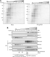
Sucrose density gradient centrifugation shows association of erlins with high molecular weight complexes. Sucrose density gradient centrifugation was performed on total lysates from HeLa cells or 3T3 cells stably expressing pLPCX vector only, E1HA, or E2HA. Cells were lysed in 1% Triton X-100 and fractionated by 40–5% sucrose gradient centrifugation. A, gels with gradient fractions from HeLa and 3T3 pLPCX cells were stained to visualize distribution of cellular proteins within the gradient. B, Western blot analyses of fractions: Distribution of endogenous erlin proteins within gradient was determined by probing Western blots with antibodies specific for either erlin-1 or erlin-2 (HeLa) or with PAN-erlin antibody, which detects both erlins (3T3). Ectopically expressed HA-tagged erlin-1 and -2 were detected with an HA-specific antibody. Western blots were also probed for calnexin, which localizes to the ER, but is not enriched in high density fractions. Distribution of molecular weight standards is indicated at the bottom. 440- and 669-kDa standards were not separated by this procedure and therefore concentrated in identical fractions.
FIGURE 3.
FIGURE 3.
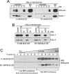
Disrupting lipid raft association of the erlins affects neither their ability to interact with each other nor their association with high molecular weight complexes. Crude membrane fractions from HeLa cells were treated with PBS only (0 mm M-β-CD) or 50 mm M-β-CD in PBS for 30 min at 37 °C and subsequently subjected to lipid raft isolation (A), immunoprecipitation (B), and sucrose gradient centrifugation (C). A, pretreated membrane fractions were lysed in 1% Triton X-100, lysate was adjusted to 40% sucrose and overlaid with a 30–5% continuous sucrose gradient. After ultracentrifugation six fractions were collected from gradient, which were further separated into soluble and insoluble subfractions. Equal parts of insoluble fractions 1–6 and soluble protein from fraction 6 were analyzed by Western blotting using the PAN-erlin antibody and a flotillin-1-specific antibody as lipid raft marker. B, immunoprecipitation of endogenous erlins was performed as in Fig. 1D. Lysate: 20% of input. C, sucrose gradient centrifugation was performed as in Fig. 2, and Western blot was probed with PAN-erlin antibody.
FIGURE 4.
FIGURE 4.
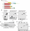
A C-terminal region of Erlin-2 between the SPFH domain and the non-conserved C terminus is required for its oligomerization and association with high molecular weight complexes and DRMs. A, schematic of full-length and truncated erlin-2 constructs used for initial characterization in this study: Transmembrane (TM) domain is shown in gray, prohibitin (PHB) domain is shown in red, red and orange boxes combined represent the SPFH domain, and red, orange, and yellow boxes combined represent HflK/C domain. The blue box indicates a C-terminal region, which shows very little conservation between erlin-1 and -2. HA and FLAG tags (green) were added to the C terminus of the erlins. Erlin-2 truncations lack various parts of the C terminus and were named N188, N227, and N305, because they consist of the N-terminal 188, 227, and 305 amino acids, respectively. B, full-length (FL) FLAG-tagged erlin-2 and HA-tagged truncated (or FL) erlin-2 were stably co-expressed in NIH-3T3 cells, and immunoprecipitations with HA- and FLAG-specific antibodies were performed. Western blots on the left show total lysate, the middle and right panels show immunoprecipitations HA- and FLAG-specific antibodies, respectively. Lanes in the middle and right panels marked with “+” show IPs with HA- or FLAG-specific antibodies, whereas lanes marked with “-” show control IPs with normal rat or mouse IgGs, respectively. Lysate: 20% of input. C, sucrose gradient centrifugation was performed on 3T3 cells stably expressing truncated or FL erlin-2HA, as described for Fig. 3. D, lipid raft isolation was performed on 3T3 cells stably expressing truncated or FL erlin-2HA. Equal portions of soluble (s) and raft (r) fractions were analyzed by Western blotting to determine lipid raft association of erlin-2 truncation mutants. Flotillin-1 was used as a marker for lipid raft fractions.
FIGURE 5.
FIGURE 5.
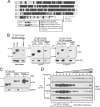
Residue Phe-305 within a short hydrophobic stretch is dispensable for oligomerization and lipid raft association but is required for high molecular weight complex association of erlin-2. A, ClustalW alignment of amino acid sequences of erlin-1 and -2 (modified from reference 6); the small gray box highlights residues 301–310, which are magnified in the large gray box. Hydrophobic residues between 301 and 306 are marked as bold letters, and underlined residues are included in erlin-2 N305 truncation mutant. The black box contains a list of additional erlin-2 mutants. B, testing new erlin-2HA mutants for oligomerization using the immunoprecipitation approach described for Fig. 4B. On α-HA, a Western blot shows total lysate (top left), and 10× the amount of protein was loaded onto the gel for IPMFM→A compared with N300 and F305A. Longer exposure times are shown for IP blots of IPMFM→A mutant, due to low expression levels of this mutant in that specific cell line. Lysate: 10% of input. C, testing lipid raft association of new erlin-2HA mutants using same approach as in Fig. 4D. D, association of new erlin-2HA mutants was examined using sucrose gradient centrifugation, as described for Fig. 4C. Western blot of FL E2HA gradient fractions was included for comparison.

References

    1. Tavernarakis, N., Driscoll, M., and Kyrpides, N. C. (1999) Trends Biochem. Sci. 24 425-427 - PubMed
    1. Browman, D. T., Hoegg, M. B., and Robbins, S. M. (2007) Trends Cell Biol. 17 394-402 - PubMed
    1. Morrow, I. C., and Parton, R. G. (2005) Traffic 6 725-740 - PubMed
    1. Rivera-Milla, E., Stuermer, C., and Malaga-Trillo, E. (2006) Cell. Mol. Life Sci. 63 343-357 - PMC - PubMed
    1. Huber, T. B., Schermer, B., Muller, R. U., Hohne, M., Bartram, M., Calixto, A., Hagmann, H., Reinhardt, C., Koos, F., Kunzelmann, K., Shirokova, E., Krautwurst, D., Harteneck, C., Simons, M., Pavenstadt, H., Kerjaschki, D., Thiele, C., Walz, G., Chalfie, M., and Benzing, T. (2006) Proc. Natl. Acad. Sci. U. S. A. 103 17079-17086 - PMC - PubMed

Publication types

LinkOut - more resources