. 2009 Apr 3;284(14):9498-512.
doi: 10.1074/jbc.M806136200. Epub 2009 Jan 8.

Rational design of alpha-conotoxin analogues targeting alpha7 nicotinic acetylcholine receptors: improved antagonistic activity by incorporation of proline derivatives

Affiliations

Rational design of alpha-conotoxin analogues targeting alpha7 nicotinic acetylcholine receptors: improved antagonistic activity by incorporation of proline derivatives

Christopher Armishaw et al. J Biol Chem. .

Abstract

Nicotinic acetylcholine receptors (nAChRs) are ligand-gated ion channels that belong to the superfamily of Cys loop receptors. Valuable insight into the orthosteric ligand binding to nAChRs in recent years has been obtained from the crystal structures of acetylcholine-binding proteins (AChBPs) that share significant sequence homology with the amino-terminal domains of the nAChRs. alpha-Conotoxins, which are isolated from the venom of carnivorous marine snails, selectively inhibit the signaling of neuronal nAChR subtypes. Co-crystal structures of alpha-conotoxins in complex with AChBP show that the side chain of a highly conserved proline residue in these toxins is oriented toward the hydrophobic binding pocket in the AChBP but does not have direct interactions with this pocket. In this study, we have designed and synthesized analogues of alpha-conotoxins ImI and PnIA[A10L], by introducing a range of substituents on the Pro(6) residue in these toxins to probe the importance of this residue for their binding to the nAChRs. Pharmacological characterization of the toxin analogues at the alpha(7) nAChR shows that although polar and charged groups on Pro(6) result in analogues with significantly reduced antagonistic activities, analogues with aromatic and hydrophobic substituents in the Pro(6) position exhibit moderate activity at the receptor. Interestingly, introduction of a 5-(R)-phenyl substituent at Pro(6) in alpha-conotoxin ImI gives rise to a conotoxin analogue with a significantly higher binding affinity and antagonistic activity at the alpha(7) nAChR than those exhibited by the native conotoxin.


Figures

FIGURE 1.
FIGURE 1.
A, three-dimensional NMR structure of α-conotoxin ImI (75); B, x-ray crystal structure of α-conotoxin PnIA (76) showing the consensus fold of α-conotoxins. The Pro6 residue is highlighted in yellow. C, three-dimensional crystal structure of α-conotoxin ImI; D, [A10L]-PnIA bound to Ac-AChBP showing the interactions of these ligands to AChBP. The conotoxins are shown in yellow, and the two chains of AChBP are shown in gray and blue, respectively. Residues that form the hydrophobic binding pocket are indicated.
FIGURE 2.
FIGURE 2.
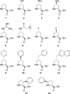
Proline derivatives used in this study. 1, l-proline; 2, 4-(R)-hydroxy-l-proline, [4-(R)-OH]; 3, 4-(R)-amino-l-proline, [4-(R)-NH2]; 4, 4-(S)-amino-l-proline, [4-(S)-NH2]; 5, 4-(R)-guanidino-l-proline, [4-(R)-Gn]; 6, 4-(R)-betainamidyl-l-proline, [4-(R)-Bet]; 7, 4-(R)-fluoro-l-proline, [4-(R)-F]; 8, 4-(S)-fluoro-l-proline, [4-(S)-F]; 9, 4-(R)-phenyl-l-proline, [4-(R)-Ph]; 10, 4-(S)-phenyl-l-proline, [4-(S)-Ph]; 11, 4-(R)-1-naphthylmethyl-l-proline, [4-(R)-Nap]; 12, 4-(R)-benzyl-l-proline, [4-(R)-Bzl]; 13, 3-(S)-phenyl-l-proline, [3-(S)-Ph]; 14, 5-(R)-phenyl-l-proline, [5-(R)-Ph].
FIGURE 3.
FIGURE 3.
Circular dichroism spectra of α-conotoxin Pro6 analogues. The % helix is indicated in the legend and was estimated as described by Woody (66). A, ImI analogues incorporating polar substituents; B, ImI analogues incorporating electronegative substituents on Pro6; C, ImI analogues incorporating aromatic substituents; D, PnIA[A10L] analogues incorporating polar substituents; E, PnIA[A10L] analogues incorporating electronegative substituents on Pro6; F, PnIA[A10L] analogues incorporating aromatic substituents.
FIGURE 4.
FIGURE 4.
Three-dimensional structure of ImI[P6/5-(R)-Ph]. A, ribbon representation of the mean structure of ImI[P6/5-(R)-Ph] showing the two 310 helical regions and the two disulfides (Cys2-Cys8 and Cys3-Cys12) in ball and stick representation. B, comparison of the peptide backbone conformation of ImI[P6/5-(R)-Ph] (blue) and WT-ImI (PDB ID 1im1) (red) (68). The side chains at position 6 are also shown to illustrate the extension of the hydrophobic phenyl ring in ImI[P6/5-(R)-Ph]. C, surface representations of ImI[P6/5-(R)-Ph] (left) and WT-ImI (right). Hydrophobic residues are shown in green, polar in white, positively charged in blue, negatively charged in red, cystines in yellow, and glycine in cyan. Selected residue numbers are also shown. It is clear that the hydrophobic patch of ImI[P6/5-(R)-Ph] is significantly increased in size compared with WT-ImI.
FIGURE 5.
FIGURE 5.
Pharmacological properties of selected α-conotoxin ImI and PnIA[A10L] analogues. A, concentration-inhibition curves for WT α-conotoxin ImI and selected analogues at the α7/5-HT3A chimera in [3H]MLA binding assay (left) and at the human α7-GH3 cell line in the Fluo-4/Ca2+ assay (right). B, concentration-inhibition curves for WT α-conotoxin PnIA[A10L] and selected analogues at the α7/5-HT3A chimera in [3H]MLA binding assay (left) and at the human α7-GH3 cell line in the Fluo-4/Ca2+ assay (right). The binding and functional experiments were performed as described under “Experimental Procedures.” In the binding assay, a tracer concentration of 0.5 nm [3H]MLA was used. In the Fluo-4/Ca2+ assay, an assay concentration of 30 μm ACh was used as agonist, and the assay was performed in the presence of 100 μm genistein. The figure depicts data from single representative experiments, and error bars are omitted for reasons of clarity.
FIGURE 6.
FIGURE 6.
The α7 nAChR homology model and top scoring docking poses. A, WT α-conotoxin ImI; B, ImI[P6/4-(R)-Ph]; C, ImI[P6/4-(R)-Bzl]; D, ImI[P6/4-(R)-Nap]; E, ImI[P6/5-(R)-Ph]; F, ImI[P6/3-(S)-Ph].
FIGURE 7.
FIGURE 7.
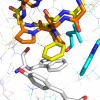
Superimposition of α-conotoxin PnIA[A10L] (orange) onto a homology model of α7 nAChR (ImI[P6/5-(R)-Ph] is shown in yellow).

References

    1. Sine, S. M., and Engel, A. G. (2006) Nature 440 455-463 - PubMed
    1. Jensen, A., Frølund, B., Liljefors, T., and Krogsgaard-Larsen, P. (2005) J. Med. Chem. 48 4705-4744 - PubMed
    1. Sher, E., Chen, Y., Sharples, T. J., Broad, L. M., Benedetti, G., Zwart, R., McPhie, G. I., Pearson, K. H., Baldwinson, T., and DeFillipi, G. (2004) Curr. Top. Med. Chem. 4 283-297 - PubMed
    1. Lukas, R. J., Changeux, J.-P., Le Novere, N., Albuquerque, E. X., Balfour, D. J. K., Berg, D. K., Bertrand, D., Chiappinelli, V. A., Clarke, P. B. S., Collins, A. C., Dani, J. A., Grady, S. R., Kellar, K. J., Lindstrom, J. M., Marks, M. J., Quik, M., Taylor, P. W., and Wonnacott, S. (1999) Pharmacol. Rev. 51 397-401 - PubMed
    1. Armishaw, C. J., and Alewood, P. F. (2005) Curr. Protein Pept. Sci. 6 221-240 - PubMed

Publication types

MeSH terms

LinkOut - more resources