. 2009 Jan 15;23(2):181-94.
doi: 10.1101/gad.1735109. Epub 2009 Jan 8.

Robust production and passaging of infectious HPV in squamous epithelium of primary human keratinocytes

Affiliations

Robust production and passaging of infectious HPV in squamous epithelium of primary human keratinocytes

Hsu-Kun Wang et al. Genes Dev. .

Abstract

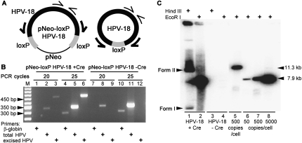
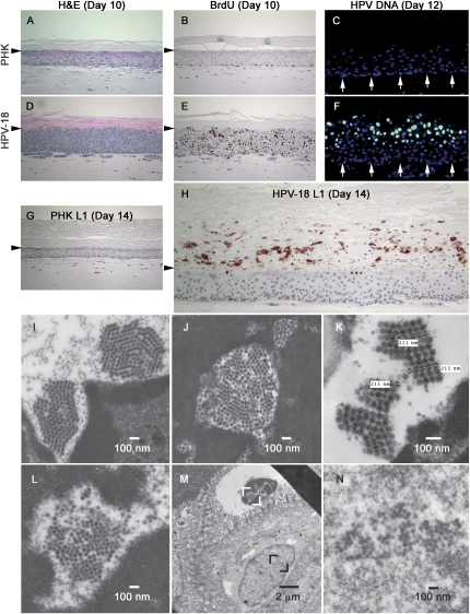
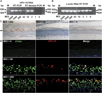
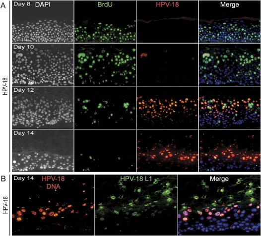
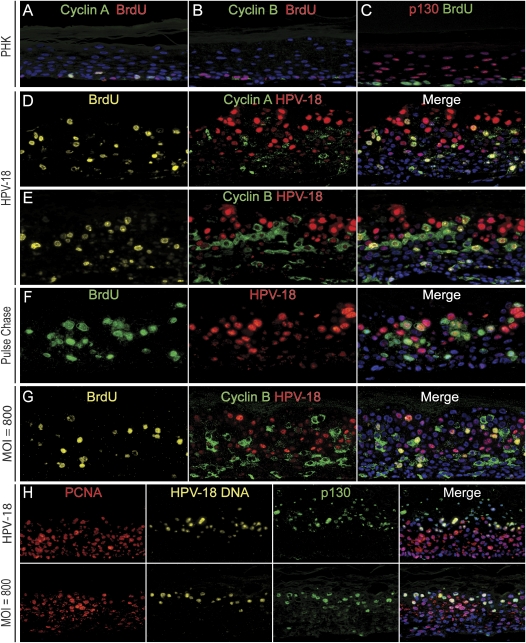
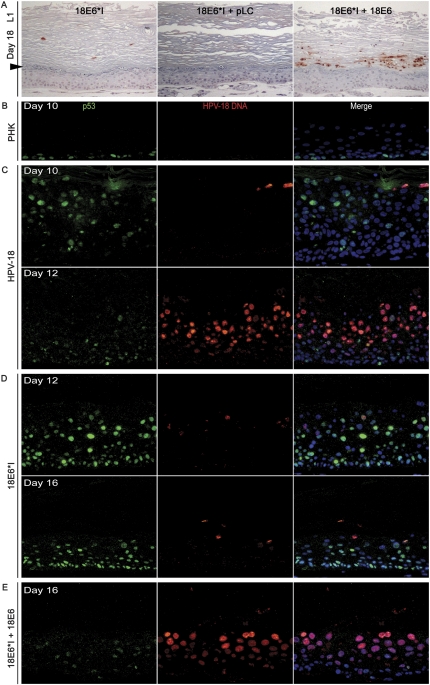
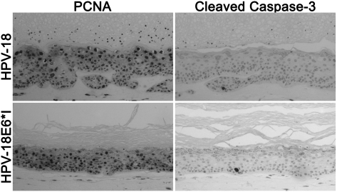
Using Cre-loxP-mediated recombination, we established a highly efficient and reproducible system that generates autonomous HPV-18 genomes in primary human keratinocytes (PHKs), the organotypic raft cultures of which recapitulated a robust productive program. While E7 promoted S-phase re-entry in numerous suprabasal differentiated cells, HPV DNA unexpectedly amplified following a prolonged G2 arrest in mid- and upper spinous cells. As viral DNA levels intensified, E7 activity diminished and then extinguished. These cells then exited the cell cycle to undergo virion morphogenesis. High titers of progeny virus generated an indistinguishable productive infection in naïve PHK raft cultures as before, never before achieved until now. An immortalization-defective HPV-18 E6 mutant genome was also characterized for the first time. Numerous cells accumulated p53 protein, without inducing apoptosis, but the productive program was severely curtailed. Complementation of mutant genomes by E6-expressing retrovirus restored proper degradation of p53 as well as viral DNA amplification and L1 production. This system will be invaluable for HPV genetic dissection and serves as a faithful ex vivo model for investigating infections and interventions.


Figures

Figure 1.
Figure 1.
HPV DNA excision and amplification. (A) Schematic diagrams depicting the parental pNeo-loxP HPV-18 and Cre-excised HPV-18 plasmids and PCR primers (thin and thick arrows) to detect total and excised HPV DNA. (B) Ethidium bromide-stained agarose gel of PCR amplification products after 20 and 25 cycles, revealing total HPV DNA (332 bp), excised HPV DNA (450 bp), or β-globin gene (275 bp) from submerged PHK cultures after transfection with pNeo-loxP HPV-18 plasmid with (lanes 1–6) or without (lanes 7–12) the nls-Cre expression plasmid. (M) DNA length markers. (C) Southern blot hybridization for HPV genomic plasmid in day-12 raft cultures. Total DNA from raft cultures of PHKs transfected with pNeo-loxP HPV-18 in the presence (lanes 1,2) or absence (lanes 3,4) of the nls-Cre expression plasmid. Length and copy number standards for EcoR I-linearized parental plasmid (lane 5) or HPV-18 genomic DNA (lanes 6–8).
Figure 2.
Figure 2.
Productive HPV-18-containing PHK raft cultures and visualization of HPV virions by TEM. (A–H) Four-micron sections of control (A–C,G) or HPV-18-containing PHK raft cultures (D–F,H) analyzed. Day-10 culture sections were stained with H&E (A,D) or probed for BrdU incorporation (reddish brown, B,E) after a 12-h exposure immediately prior to harvest. (C,F) DNA-FISH to reveal HPV-18 DNA (green) in a day-12 culture sections. Arrows point to the basal stratum. (G,H) IHC to detect the major capsid protein L1 (reddish brown) in a day-14 culture sections. Horizontal arrowheads point to the boundary between the upper cornified layer and live epithelium below. Under the epithelium is the collagen matrix. (I–N) TEM of ultrathin sections of day-14 cultures to visualize virions. (I,J) Paracrystalline arrays of mature virions in degenerated nuclei with condensed chromatins. (K) Three measurements, each across five virions in a closely packed ordered array, gave the same value of 211 nm, or 42.2 nm/virion diameter. (M) Two adjacent cells shown in low magnification. (L) The enlarged area (white brackets) in the upper cornified envelope with a degenerated nucleus. (N) The enlarged area (black brackets) in the lower, living cells with a normal nucleus.
Figure 3.
Figure 3.
Infectivity assays of HPV-18 virions in PHKs. HPV virions were titered by quantitative real-time PCR. (A) HPV cDNA detection in submerged PHK cultures after infection at the indicated MOI. A 521-bp cDNA fragment of a spliced early viral mRNA was detected by ethidium bromide staining after RT–PCR (left panel) or RT-nested PCR (right panel). (B) A 642-bp-long β-actin cDNA served as an internal control. (M) 50-bp ladder. (C) Immunohistochemical detection of the HPV major capsid protein L1 (reddish brown) in 14-d raft cultures of PHKs infected with HPV virions.(Left panel) MOI of 800 or higher. (Middle panel) MOI of 50 up to 400. An uninfected culture is shown in the right panel. Arrowheads point to the boundary between the upper cornified strata and live epithelium below. (D) Four-micron sections from the raft cultures above were probed for PCNA (Alexa Fluor 488, green) and for viral DNA (Cy3, red). Cellular DNA was revealed by DAPI (blue).
Figure 4.
Figure 4.
(A) HPV-18 DNA amplification in differentiated keratinocytes lags behind cellular DNA replication. HPV-18-containing PHK raft cultures were harvested on days 8, 10, 12, and 14, each following a 12-h incubation with BrdU. Sections were probed for HPV-18 DNA (red) and incorporated BrdU (green). Cellular DNA was revealed by DAPI (blue). (Left column) For better visualization of tissue morphology, DAPI staining is also presented in black-and-white images. (B) Detection of viral DNA (red) and the major capsid protein L1 (green) in a 14-d-old culture.
Figure 5.
Figure 5.
HPV-18 plasmid DNA amplification as it relates to the cell cycle. (A–C) Day-10 raft cultures of normal PHK raft cultures. (D–F) Day-12 raft cultures initiated from PHKs transfected with HPV-18 DNA. Sections were analyzed for cyclin A (green in A,D) or cyclin B (green, B,E), HPV-DNA FISH (red, D–F), and for BrdU (red in A,B, green in C, or yellow Alexa Fluor 647 in D,E). (F) An HPV-18-containing raft culture was pulsed with BrdU for 6 h on day 10 and then chased for 48 h prior to harvest. (G) Day 14 of PHK raft cultures infected with HPV-18 virus at MOI of 800. The section was probed for viral DNA (red), cyclin B1 (green), and BrdU (gold). (H) Patterns of PCNA (red), HPV-18 DNA (gold), and p130 (green) in day-14 raft cultures of PHKs initiated after HPV-18 DNA transfection (top row) or HPV-18 virus infection at MOI of 800 (bottom row). Cellular DNA was revealed by DAPI (blue) in all panels.
Figure 6.
Figure 6.
Complementation of HPV-18 E6*I genome by a retrovirus delivering HPV-18 URR-E6. Raft cultures were harvested over a time course. (A) IHC to detect L1 (reddish brown) on day-18 raft cultures. (Left panel) HPV-18 E6*I-containing cultures. (Middle panel) HPV-18 E6*I-containing PHKs infected with the empty pLC retrovirus. (Right panel) HPV-18 E6*I-containing PHKs trans-complemented with the pLJ HPV-18 URR-E6 retrovirus. Arrowhead points to the boundary between the upper cornified strata and live epithelium below. (B–E) Double-fluorescence detection of p53 (green) and HPV-18 DNA (red) in raft cultures. (B) Day-10 normal PHKs. (C) Day-10 and day-12 wild-type HPV-18-containing PHKs. (D) Day-12 and day-16 HPV-18 E6*I-containing PHKs. (E) Day-16 HPV-18 E6*I-containing PHKs trans-complemented with pLJ HPV-18 URR-E6. Cellular DNA was stained with DAPI (blue).
Figure 7.
Figure 7.
Widespread induction of PCNA and virtual absence of cleaved caspase 3 in raft cultures harboring the wild-type or E6*I mutant plasmid. IHC was conducted to detect PCNA (left column) or the cleaved caspase 3 (right column) in day-16 raft cultures of PHK transfected with the wild-type HPV-18 (top row) or the HPV-18 E6*I mutant (bottom row). The small dots in cornified layers represent parakeratosis.

Comment in

References

    1. Banerjee N.S., Chow L.T., Broker T.R. Retrovirus-mediated gene transfer to analyze HPV gene regulation and protein functions in organotypic ‘raft’ cultures. Methods Mol. Med. 2005;119:187–202. - PubMed
    1. Bertrand P., Saintigny Y., Lopez B.S. p53's double life: Transactivation-independent repression of homologous recombination. Trends Genet. 2004;20:235–243. - PubMed
    1. Buck C.B., Pastrana D.V., Lowy D.R., Schiller J.T. Generation of HPV pseudovirions using transfection and their use in neutralization assays. Methods Mol. Med. 2005a;119:445–462. - PubMed
    1. Buck C.B., Thompson C.D., Pang Y.Y., Lowy D.R., Schiller J.T. Maturation of papillomavirus capsids. J. Virol. 2005b;79:2839–2846. - PMC - PubMed
    1. Campo M.S. Animal models of papillomavirus pathogenesis. Virus Res. 2002;89:249–261. - PubMed

Publication types

MeSH terms

Substances