. 2009 Jan 15;23(2):195-207.
doi: 10.1101/gad.505709. Epub 2009 Jan 8.

Drosophila PTB promotes formation of high-order RNP particles and represses oskar translation

Affiliations

Drosophila PTB promotes formation of high-order RNP particles and represses oskar translation

Florence Besse et al. Genes Dev. .

Abstract

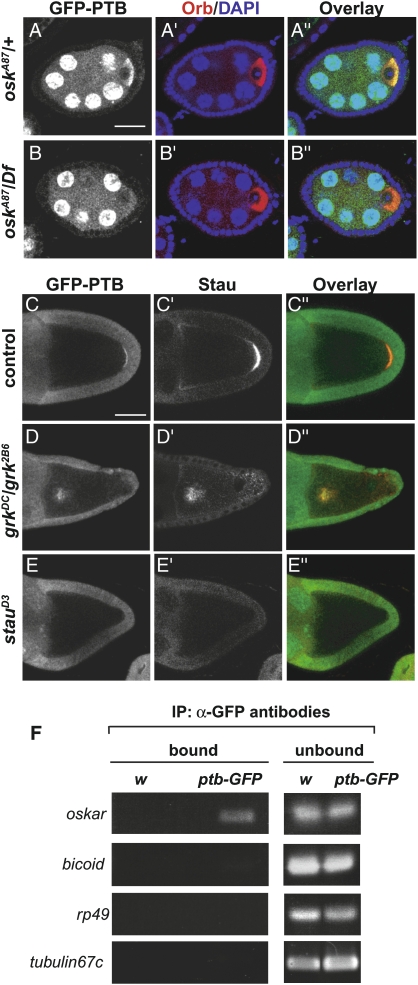
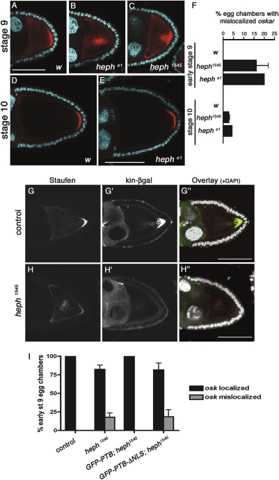
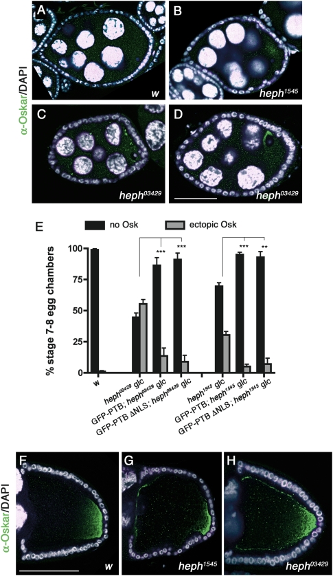
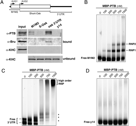
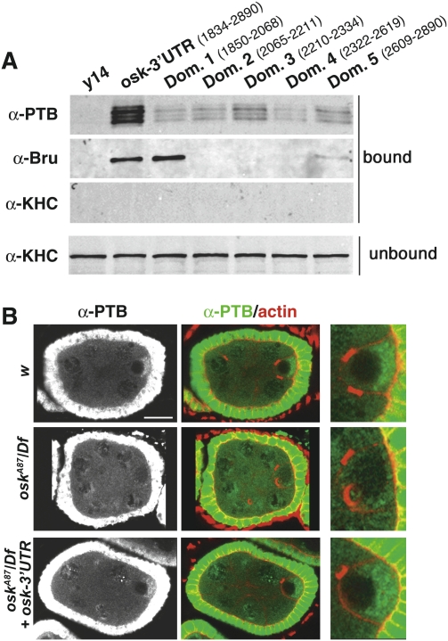
Local translation of asymmetrically enriched mRNAs is a powerful mechanism for functional polarization of the cell. In Drosophila, exclusive accumulation of Oskar protein at the posterior pole of the oocyte is essential for development of the future embryo. This is achieved by the formation of a dynamic oskar ribonucleoprotein (RNP) complex regulating the transport of oskar mRNA, its translational repression while unlocalized, and its translational activation upon arrival at the posterior pole. We identified the nucleo-cytoplasmic shuttling protein PTB (polypyrimidine tract-binding protein)/hnRNP I as a new factor associating with the oskar RNP in vivo. While PTB function is largely dispensable for oskar mRNA transport, it is necessary for translational repression of the localizing mRNA. Unexpectedly, a cytoplasmic form of PTB can associate with oskar mRNA and repress its translation, suggesting that nuclear recruitment of PTB to oskar complexes is not required for its regulatory function. Furthermore, PTB binds directly to multiple sites along the oskar 3' untranslated region and mediates assembly of high-order complexes containing multiple oskar RNA molecules in vivo. Thus, PTB is a key structural component of oskar RNP complexes that dually controls formation of high-order RNP particles and translational silencing.


Figures

Figure 1.
Figure 1.
Distribution of PTB during Drosophila oogenesis. (A,B) GFP expression pattern in an ovariole (A) and a stage 10 egg chamber (B) from homozygous GFP-PTB protein-trap females. An ovariole contains egg chambers of different developmental stages, from stage 1 in the germarium, until stage 14 (not shown). The oocyte and its 15 sibling nurse cells develop as a syncytium. The 16-cell germline cyst is surrounded by a layer of somatic epithelial cells. Egg chambers are oriented anterior to the left, posterior to the right. The oocyte, whose size dramatically increases as oogenesis proceeds, is the most posterior cell of the germline cyst. Bars, 60 μm. (C) Schematic representation of Drosophila PTB domain organization indicating the percentage of amino acid identity of the RRM domains compared with PTB orthologs, as well as the insertion site of the GFP sequence (red triangle) in the protein-trap line. (D) Western blot of ovarian extracts from w females (first lane), females heterozygous (protein-trap/+; second lane) or homozygous (protein-trap; third lane) for the protein-trap insertion, probed with rat anti-PTB antibodies. (E–G) w stage 5 (E), stage 8, (F) and stage 9 (G) egg chambers triple-stained with anti-PTB antibodies (top panel, red in the overlay), anti-Staufen antibodies (middle panel, blue in the overlay), and phalloidin (green in the overlay). Note that PTB is expressed both in the germline and in the somatic epithelium. In contrast, Staufen exclusively accumulates in the oocyte. Bar, 60 μm.
Figure 2.
Figure 2.
PTB colocalizes and associates with oskar mRNA. (A,B) Stage 5 egg chambers from oskA87/+ (A) and oskA87/Df(3R)pXT03 (B) females expressing GFP-PTB constructs under the control of the mat-α4-tub promoter, and triple-stained for GFP (green in the overlay), DNA (blue, DAPI), and the oocyte cytoplasm marker Orb (red). Bar, 15 μm. (C–E) Distribution of the GFP-PTB protein-trap fusion in wild-type (C), grk2E12/grk2B6 (D), and stauD3 (E) oocytes. (Left) GFP signal. (Middle) Staufen protein. (Right) Overlay. Bar, 60 μm. (F, left panel) RT–PCR amplification of mRNAs recovered in fractions immunoprecipitated from GFP-PTB protein-trap or w control ovarian extracts using anti-GFP antibodies. (Right panel) Amplifications from unbound fractions are used as controls.
Figure 3.
Figure 3.
PTB modulates oskar mRNA localization and oocyte MT polarity. (A–E) Double staining of early stage 9 (A–C) or stage 10 (D,E) oocytes with anti-Staufen antibodies (red) and DAPI (cyan). (A,D) w. (B,E) OvoD-selected hephe1 germline clone. (C) OvoD-selected heph1545 germline clone. Bars, 60 μm. (F) Percentage of early stage 9 or stage 10 oocytes in which Staufen was mislocalized (at least 56 oocytes were scored per genotype). (G,H) Stage 9 oocytes heterozygous (G) and homozygous (H) for heph1545, and triple-stained with anti-Staufen (G,H, red in the overlay), anti-βgal (G′,H′, green in the overlay), and DAPI (white in the overlay). Bar, 60 μm. (I) Graph showing the percentages of early stage 9 oocytes with mislocalized oskar mRNA in different mutant and rescue contexts. GFP-PTB and GFP-PTBΔNLS constructs were expressed under the control of the germline-specific promoter mat-α4-tub. Expression levels of the insertions chosen for the rescue experiments were similar, and comparable with endogenous levels (data not shown). The number of egg chambers counted per genotype is control (n = 62); heph1545 (n = 86); GFP-PTB; heph1545 (n = 56); GFP-PTBΔNLS; heph1545 (n = 56).
Figure 4.
Figure 4.
ptb mutant oocytes display ectopic Oskar protein accumulation. (A–D) w (A), heph1545 (B), and heph03429 (C,D) stage 7 egg chambers double-stained with anti-Oskar antibodies (green) and DAPI (white). Bar, 30 μm. The different patterns of ectopic Oskar accumulation reflect the dynamic distribution of oskar mRNA at this stage, which is not altered in ptb mutant oocytes compared with wild-type oocytes (data not shown). (E) Graph showing the percentage of stage 7 oocytes with premature Oskar translation in different mutant and rescue backgrounds (see the legend for Fig. 3I for a description of the rescue constructs). (**) P < 0.005; (***) P < 0.001 in a χ2 test. (F–H) w (F), heph1545 (G), and heph03429 (H) stage 10 oocytes double-stained with anti-Oskar antibodies (green) and DAPI (white). Bar, 60 μm.
Figure 5.
Figure 5.
Characterization of PTB binding to oskar RNA. (A) RNA affinity pull-down assay using biotinylated RNAs covering the entire oskar mRNA, and w ovarian extracts. The coding sequence of y14 was used as an unrelated RNA control. (Top) Diagram showing the three different probes used in the assay. Proteins in the bound or unbound fractions were visualized after Western blot analysis using the indicated antibodies. Bruno exclusively binds to the oskar 3′UTR, whereas Khc, a component of the oskar transport machinery without RNA-binding capacity, is not detected in the precipitated fraction. (B–D) EMSA analysis using 50 nM fluorescently labeled oskar M1M2 region (B), oskar 3′UTR (C), or y14 coding sequence (D) in the presence of increasing amounts of MBP-PTB (from 0 to 1500 nM) and of 5 μM of tRNA. The position of the unbound RNA (Free-RNA) and of the discrete RNP complexes is marked on the left and right, respectively.
Figure 6.
Figure 6.
PTB binds to multiple sites along the entire oskar 3′UTR. (A) Affinity pull-down assays comparing the binding of PTB to the whole oskar 3′UTR and to individual 3′UTR subdomains (see Supplemental Fig. S5). The conditions used were similar to those described in Figure 5A. (B) PTB (green) and actin (red) staining showing the accumulation of PTB in the cytoplasm of stage 6 w oocytes, or oocytes either lacking endogenous oskar mRNA (oskA87/Df) or expressing full-length oskar 3′UTR (see the Materials and Methods for complete genotypes). Bar, 15 μm.
Figure 7.
Figure 7.
PTB promotes copackaging of oskar mRNA molecules in vivo. In situ hybridization of heterozygous (A,B) and homozygous (C,D) heph1545 late stage 9 egg chambers expressing a UAS-EGFP-osk 3′UTR construct under the control of the mat-α4-tub-Gal4 driver. (A,C) Egg chambers double-stained for EGFP transcripts (red) and DAPI (white). Images A and C were taken using identical settings. (B,D) Egg chambers double-stained for endogenous oskar transcripts (green) and DAPI (white). Endogenous oskar was detected using a probe exclusively covering the coding region. Pictures B and D were taken using identical settings. Bar, 60 μm. (E) Quantification of the in situ hybridization signal detected at the posterior pole of late stage 9 oocytes. Intensity values obtained using ImageJ are expressed as a percentage of control oocytes. (***) P < 0.001 in a Student's t-test. (F) Expression levels of rp49, endogenous oskar, and EGFP-oskar 3′UTR transcripts in both heterozygous and homozygous heph1545 backgrounds, as measured by semiquantitative RT–PCR. (G,H) Electron micrographs of w (G) and heph03429 (H) early stage 9 oocytes hybridized with an oskar antisense probe. oskar mRNA was detected using 10-nm gold particles, and the central region of the oocyte is shown. Note that in heph03429 oocytes, the slightly electron-dense oskar RNP complexes are smaller and more numerous. Bar, 150 nm.

Comment in

References

    1. Auweter S.D., Allain F.H. Structure–function relationships of the polypyrimidine tract binding protein. Cell. Mol. Life Sci. 2008;65:516–527. - PMC - PubMed
    1. Besse F., Mertel S., Kittel R.J., Wichmann C., Rasse T.M., Sigrist S.J., Ephrussi A. The Ig cell adhesion molecule Basigin controls compartmentalization and vesicle release at Drosophila melanogaster synapses. J. Cell Biol. 2007;177:843–855. - PMC - PubMed
    1. Bonin C.P., Mann R.S. A piggyBac transposon gene trap for the analysis of gene expression and function in Drosophila . Genetics. 2004;167:1801–1811. - PMC - PubMed
    1. Chang J.S., Tan L., Schedl P. The Drosophila CPEB homolog, Orb, is required for Oskar protein expression in oocytes. Dev. Biol. 1999;215:91–106. - PubMed
    1. Chekulaeva M., Hentze M.W., Ephrussi A. Bruno acts as a dual repressor of oskar translation, promoting mRNA oligomerization and formation of silencing particles. Cell. 2006;124:521–533. - PubMed

Publication types

MeSH terms

LinkOut - more resources