. 2009 Feb 26;113(9):1909-18.
doi: 10.1182/blood-2008-09-178459. Epub 2009 Jan 8.

Human erythrocytes bind and inactivate type 5 adenovirus by presenting Coxsackie virus-adenovirus receptor and complement receptor 1

Affiliations

Human erythrocytes bind and inactivate type 5 adenovirus by presenting Coxsackie virus-adenovirus receptor and complement receptor 1

Robert C Carlisle et al. Blood. .

Abstract

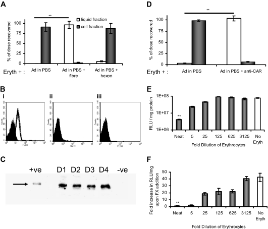
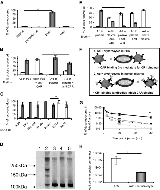
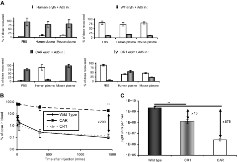
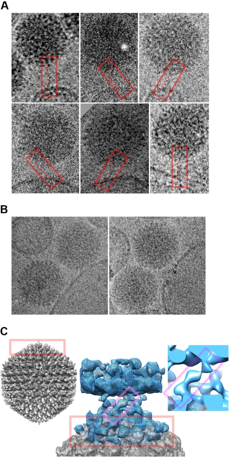
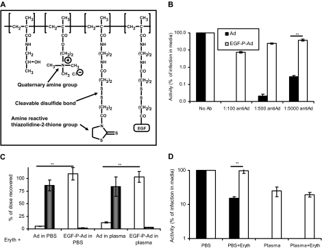
Type 5 adenovirus (Ad5) is a human pathogen that has been widely developed for therapeutic uses, with only limited success to date. We report here the novel finding that human erythrocytes present Coxsackie virus-adenovirus receptor (CAR) providing an Ad5 sequestration mechanism that protects against systemic infection. Interestingly, erythrocytes from neither mice nor rhesus macaques present CAR. Excess Ad5 fiber protein or anti-CAR antibody inhibits the binding of Ad5 to human erythrocytes and cryo-electron microscopy shows attachment via the fiber protein of Ad5, leading to close juxtaposition with the erythrocyte membrane. Human, but not murine, erythrocytes also present complement receptor (CR1), which binds Ad5 in the presence of antibodies and complement. Transplantation of human erythrocytes into nonobese diabetic/severe combined immunodeficiency mice extends blood circulation of intravenous Ad5 but decreases its extravasation into human xenograft tumors. Ad5 also shows extended circulation in transgenic mice presenting CAR on their erythrocytes, although it clears rapidly in transgenic mice presenting erythrocyte CR1. Hepatic infection is inhibited in both transgenic models. Erythrocytes may therefore restrict Ad5 infection (natural and therapeutic) in humans, independent of antibody status, presenting a formidable challenge to Ad5 therapeutics. "Stealthing" of Ad5 using hydrophilic polymers may enable circumvention of these natural virus traps.


Figures

Figure 1
Figure 1
Ad5 is inactivated by binding to human erythrocytes via the CAR. (A) Fresh washed human erythrocytes were incubated with Ad5 in the presence or absence of fiber or hexon. Ad5 erythrocyte binding was assessed by separating the liquid (□) and erythrocyte (■) fractions by centrifugation and performing quantitative PCR specific for the Ad5 genome on each fraction. Data are represented as the percentage of the total input dose recovered; n = 4, SEM shown. **P < .005. (B) Human erythrocytes from 4 donors (i) or erythrocytes from C57BL/6 mice (ii) or rhesus macaques (iii) were analyzed for CAR presentation using the anti-CAR primary antibody RmcB, secondary antibody R0480 (Dako Denmark), and flow cytometric analysis. (i) Filled peak represents isotype control (antibody W6/32); empty peak, donors 1 to 4 with anti-CAR. (ii,iii) Filled peak represents isotype control; empty peak, + anti-CAR (data are superimposed). (C) Human erythrocytes from 4 donors (D1-D4) were treated to produce hemoglobin-free ghosts and then analyzed by SDS-PAGE/Western blotting using 100 ng/lane, primary antibody 15405 (Santa Cruz Biotechnology), and secondary antibody W4011 (Promega). Positive control (+ve) indicates mouse liver lysate (10 ng); negative control, mouse A9 cells (∼ 100 ng). (D) Human erythrocytes in PBS were incubated with anti-CAR antibody (RmcB) before Ad5 addition. After separation of the liquid (□) and cell fraction (■) by centrifugation, quantification of each fraction was achieved by quantitative PCR; n = 4, SEM shown. **P < .005. (E) A549 cells were incubated with Ad5 in the presence of serially diluted erythrocytes for 90 minutes at 37°C; after thorough washing in PBS and the addition of fresh media, the A549 cells were returned to the incubator and luciferase expression was measured 24 hours later; N = 4, SEM shown. **P < .005. (F) SKOV-3 cells were incubated with Ad5 in the presence or absence of physiologic FX levels (8 μg/mL) and serially diluted erythrocytes in PBS, for 90 minutes at 37°C; after thorough washing in PBS and the addition of fresh media, cells were returned to the incubator and luciferase expression was measured 24 hours later. The fold increase on FX addition was calculated by dividing the value achieved with FX by that achieved without FX, and plotted for each erythrocyte dilution; N = 4, SEM shown. **P < .005.
Figure 2
Figure 2
Human plasma mediates binding of Ad5 to erythrocytes via the complement cascade and CR1. (A) Ad5 was incubated with whole fresh blood and cell fractionation using CPT vacutainers (BD Biosciences) and erythrocyte lysis was performed; the amount of Ad5 associated with each fraction was then quantified by quantitative PCR; N = 4 separate donors, SEM shown. **P < .005. (B) Erythrocytes were isolated and washed before resuspension in either PBS or neutralizing plasma. Erythrocytes were preincubated or not with anti-CAR antibody (RmcB) and then incubated with Ad5; after separation of the liquid (□) and cell fraction (■) by centrifugation, quantification was achieved by quantitative PCR; n = 4, SEM shown, **P < .005. (C) Erythrocytes were thoroughly washed in PBS and resuspended in a variety of plasmas. Ad5 was added and after incubation, the liquid (□) and erythrocyte (■) fractions were separated by centrifugation and analyzed by quantitative PCR for Ad5 genome content (see “Quantitation of Ad5 binding to erythrocytes by real-time (quantitative) PCR”); N = 4, SEM shown. **P < .005. (D) Western blot analysis using anti-C3 antibody was performed to detect the formation of covalent C3-Ad5 adducts: lane 1 indicates Ad5; lane 2, Ad5 + heparin plasma; lane 3, Ad5 + ethylenediaminetetraacetic acid plasma; lane 4, heparin plasma; lane 5, ethylenediaminetetraacetic acid plasma. (E) In human plasma, inhibitors of the binding of complement to CR1 inhibit the association of Ad5 with erythrocytes. Ad5 was added to erythrocytes in plasma in the presence or absence of antibodies against CR1 or C1q or to plasma that had been pretreated with CVF or heat treated to 56°C for 30 minutes. After fractionation of erythrocytes and plasma, Ad5 genome content of each fraction (□ represents plasma; ■, cells) was quantified by quantitative PCR; N = 4, SEM shown. **P < .005. (F) Schematic representing the binding of Ad5 to erythrocytes in PBS or plasma. In PBS, Ad5 binds erythrocytes via CAR but cannot bind via CR1 because of an absence of complement. In neutralizing plasma, Ad5 binds via CR1 but cannot bind to CAR because the epitopes of fiber protein responsible for such binding are covered by neutralizing antibodies. (G) The presence of human erythrocytes in NOD-SCID mice alters Ad5 circulation kinetics. Ad5 was administered intravenously to NOD-SCID mice transplanted with a 10% vol/vol total blood volume of washed human erythrocytes. At defined time points, blood was sampled, separated into plasma and cell fractions, and assayed for Ad5 content by quantitative PCR. Black line/triangle represents dose recovered from the cell fraction of mice treated with human erythrocytes; black dashed line/square, cell fraction from mice that did not receive human erythrocytes; gray line/triangle, plasma fraction from mice that received human erythrocytes; gray dashed line/square, plasma fraction from mice that did not receive human erythrocytes; N = 3, SD shown. **P < .005. (H) The presence of human erythrocytes in NOD-SCID mice inhibits deposition of an intravenously administered Ad5 dose within subcutaneous xenograft tumors. HT29 cells were implanted into NOD-SCID mice and allowed to establish. Mice were injected with Ad5 in the presence or absence of 10% (vol/vol) total blood volume of fresh washed human erythrocytes. After 24 hours, tumors were harvested and assessed for virus genome content. Grubbs outlier test was applied to remove statistical outliers, leaving N = 4, SD shown. *P < .05.
Figure 3
Figure 3
Erythrocyte binding, circulation kinetics, and infection profiles are altered in CAR and CR1 transgenic mice. (A) Erythrocytes were isolated from human donors (i), WT mice (ii), CAR mice (iii), or CR1 mice (iv), and after washing were resuspended in PBS, human plasma, or mouse plasma. Ad5 was added and after incubation, liquid (□) and erythrocyte (■) fractions were separated and assayed for Ad5 genome content; N = 4, SEM shown. (B) The circulation kinetics of Ad5 are altered in CAR but not CR1 transgenic mice. Ad5 was injected intravenously into WT, CAR, or CR1 mice and blood sampled at 10, 30, 360, or 1440 minutes. Samples were assayed for Ad5 genome content by quantitative PCR; N = 3, SD shown. **P < .005. (C) The livers from the mice injected in panel B were harvested at 24 hours and after homogenization were assayed for reporter gene expression; N = 3, SD shown. **P < .005.
Figure 4
Figure 4
The binding of Ad5 to erythrocytes can be visualized by cryo-electron microscopy. Erythrocyte ghosts were exposed to Ad5 for 30 minutes before fixation and imaging. (A) Gallery of 6 images of Ad5 interacting peripherally with human erythrocyte ghost membranes in PBS. In each case, the tubular density visible for the pentagonal vertex fiber is boxed in red, with a series of points marking the flexible path of the fiber from the Ad5 surface above to the membrane below. (B) Images of Ad5 and ghost membranes in close association in PBS. (C) Central image shows a reconstruction of the close interaction of Ad5 with the human ghost membrane. The planar membrane (blue density mesh) forms the top part of the image, and the curved pentagonal vertex of the Ad5 (gray density surface) the bottom portion. They are linked by a crown of obliquely oriented features. The Ad5 portion of the image is superimposed with the equivalent portion of a whole-Ad5 reconstruction, for comparison. The whole Ad5 is shown to the left (gray density), in the same orientation as in the middle panel, with equivalent portions in the 2 boxed in red. A closeup of one connection is shown to the right, and equivalent portions of it and the central image are boxed in magenta.
Figure 5
Figure 5
Polymer “stealthing” can prevent unwanted binding of Ad5 to human erythrocytes. (A) Representation of HPMA-EGF used to modify Ad5. (B) Comparison of normal and EGF-mediated infection in neutralizing plasma. Ad5 or EGF-P-Ad5 was incubated with dilutions of neutralizing antisera and then added to a monolayer of A431 cells; after 90 minutes, media was removed and washing performed in PBS; and after 24 hours, luciferase expression was analyzed. (C) Ad5 or EGF-P-Ad5 was incubated with washed erythrocytes suspended in PBS/1% BSA or whole fresh human plasma. After incubation, erythrocyte and liquid fractions were separated and assayed for Ad5 genome content as described in “Quantitation of Ad5 binding to erythrocytes by real-time (quantitative) PCR” (□ represents liquid fraction; ■, cell fraction). (D) Comparison of normal and EGF-mediated infection in presence of human erythrocytes. A431 cells were infected with Ad5 or EGF-P-Ad5 in the presence of a 1 in 5 dilution of erythrocytes suspended in PBS or plasma. After 90 minutes, media was removed and thorough washing in PBS performed; 24 hours later, luciferase expression was analyzed. ■ represent Ad5; □, EGF-P-Ad. (B-D) N = 4, SEM shown. **P < .005.

References

    1. Hayashi S, Hogg JC. Adenovirus infections and lung disease. Curr Opin Pharmacol. 2007;7:237–243. - PMC - PubMed
    1. Parker AL, Waddington SN, Nicol CG, et al. Multiple vitamin K-dependent coagulation zymogens promote adenovirus-mediated gene delivery to hepatocytes. Blood. 2006;108:2554–2561. - PubMed
    1. Kalyuzhniy O, Di Paolo NC, Silvestry M, et al. Adenovirus serotype 5 hexon is critical for virus infection of hepatocytes in vivo. Proc Natl Acad Sci U S A. 2008;105:5483–5488. - PMC - PubMed
    1. Waddington SN, McVey JH, Bhella D, et al. Adenovirus serotype 5 hexon mediates liver gene transfer. Cell. 2008;132:397–409. - PubMed
    1. Peng Z. Current status of gendicine in China: recombinant human Ad-p53 agent for treatment of cancers. Hum Gene Ther. 2005;16:1016–1027. - PubMed

Publication types

MeSH terms

Substances