

Premature aging of T cells is associated with faster HIV-1 disease progression
- PMID: 19131896
- PMCID: PMC2767229
- DOI: 10.1097/QAI.0b013e3181926c28
Premature aging of T cells is associated with faster HIV-1 disease progression
Abstract
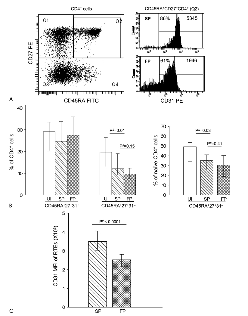
Objective: To determine if untreated HIV-1 infection and progression is associated with premature aging of memory CD8 and CD4 T cells and naive CD4 T cells.
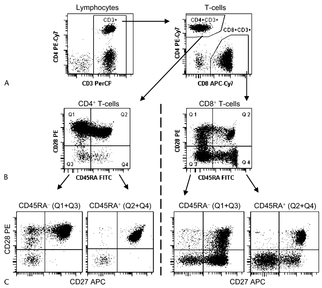
Methods: Twenty HIV-1-infected fast progressors and 40 slow progressors were included in our study, using risk set sampling. The expression of cell surface markers reflecting the differentiation stages of lymphocytes was measured using flow cytometry analyses performed on cryopreserved peripheral blood mononuclear cells.
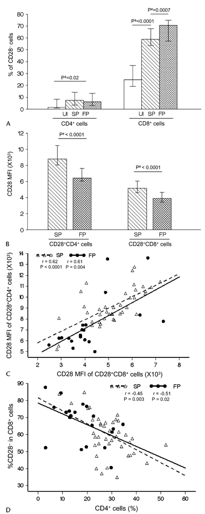
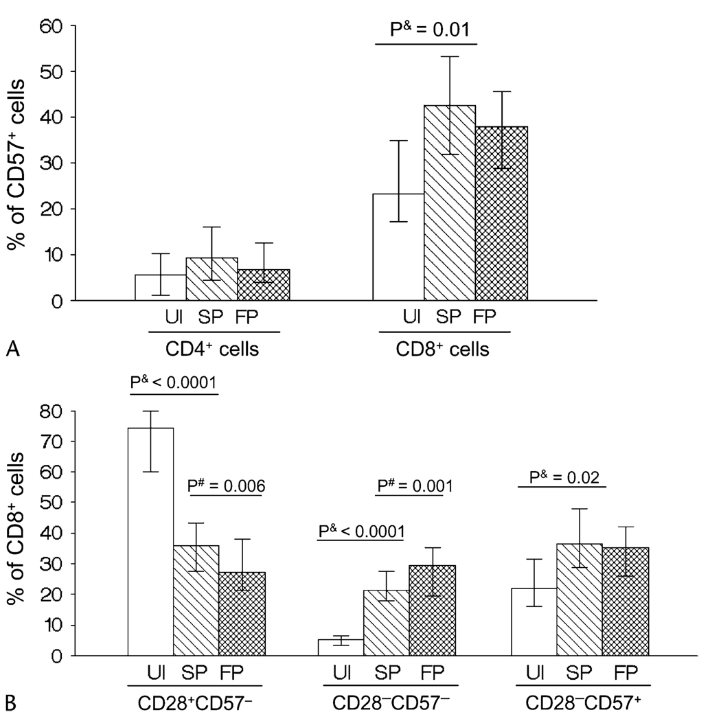
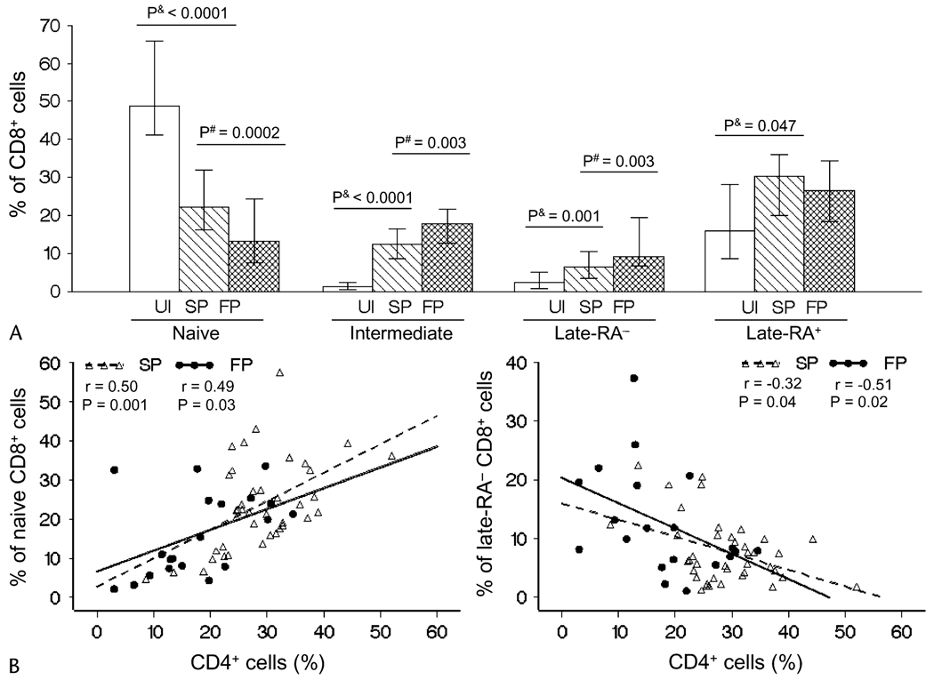
Results: We found that HIV-1 disease progression is associated with a decreased CD28 median florescence intensity on CD4 and CD8 T cells; an increased proportion of intermediate- and late-differentiated CD8 T cells and a decreased CD31 median florescence intensity on naive CD4 T cells of recent thymic origin. A selective depletion of peripherally expanded naive CD4 T cells was found to be associated with HIV-1 infection but not with HIV-1 disease progression.
Conclusions: The overall change during HIV-1 infection and progression is associated with a shift in the T-cell population toward an aged conformation, which may be further compromised by impaired renewal of the less-differentiated CD4 T-cell population. Our results suggest that HIV-1 infection induces an accelerated aging of T lymphocytes, which is associated with the clinical progression to AIDS and death.
Figures
References
-
- Appay V, Rowland-Jones SL. Premature ageing of the immune system: the cause of AIDS? Trends Immunol. 2002;23:580–585. - PubMed
-
- Effros RB. Impact of the Hayflick Limit on T cell responses to infection: lessons from aging and HIV disease. Mech Ageing Dev. 2004;125:103–106. - PubMed
-
- Pawelec G, Effros RB, Caruso C, et al. T cells and aging (update february 1999) Front Biosci. 1999;4:D216–D269. - PubMed
-
- Ho DD, Neumann AU, Perelson AS, et al. Rapid turnover of plasma virions and CD4 lymphocytes in HIV-1 infection. Nature. 1995;373:123–126. - PubMed
-
- Perillo NL, Walford RL, Newman MA, et al. Human T lymphocytes possess a limited in vitro life span. Exp Gerontol. 1989;24:177–187. - PubMed
Publication types
MeSH terms
Grants and funding
LinkOut - more resources
Full Text Sources
Medical
Research Materials
