. 2009;4(1):31-6.
doi: 10.1038/nprot.2008.214.

Immunodesign of experimental sepsis by cecal ligation and puncture

Affiliations

Immunodesign of experimental sepsis by cecal ligation and puncture

Daniel Rittirsch et al. Nat Protoc. 2009.

Abstract

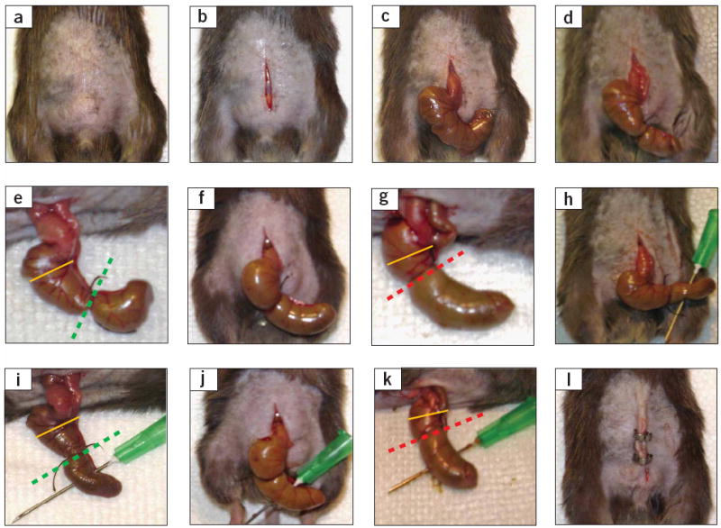
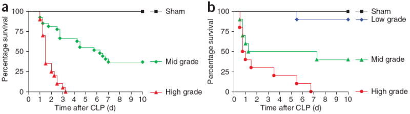
Sepsis remains a prevalent clinical challenge and the underlying pathophysiology is still poorly understood. To investigate the complex molecular mechanisms of sepsis, various animal models have been developed, the most frequently used being the cecal ligation and puncture (CLP) model in rodents. In this model, sepsis originates from a polymicrobial infectious focus within the abdominal cavity, followed by bacterial translocation into the blood compartment, which then triggers a systemic inflammatory response. A requirement of this model is that it is performed with high consistency to obtain reproducible results. Evidence is now emerging that the accompanying inflammatory response varies with the severity grade of sepsis, which is highly dependent on the extent of cecal ligation. In this protocol, we define standardized procedures for inducing sepsis in mice and rats by applying defined severity grades of sepsis through modulation of the position of cecal ligation. The CLP procedure can be performed in as little as 10 min for each animal by an experienced user, with additional time required for subsequent postoperative care and data collection.


Figures

Figure 1
Figure 1
Critical steps in the CLP procedure in mice. (a) Disinfection of the abdominal area after shaving (Step 4). (b) Skin midline incision (Step 6). (c) Exposure of the cecum, which can be mostly found in the lower left area of the abdominal cavity (Step 8). (d,e) Ligation of the cecum at designated positions as the major determinant of sepsis severity (Step 10). For the induction of mid-grade sepsis resulting in survival rates of ~ 40%, the cecum is ligated (indicated by dotted green line) at half the distance between distal pole and the base of the cecum (black dotted line). (f,g) High-grade sepsis (100% lethality) comprises ligation of 75% of the cecum (dotted red line). The basis of the cecum is indicated by the yellow line. (h,i) Cecal puncture (‘through-and-through’) from mesenteric toward antimesenteric direction after medium ligation (Step 11). (j,k) Needle puncture of the cecum under the conditions of high-grade sepsis (large ligation; Step 11). (l) Wound closure by applying simple running sutures to the abdominal musculature and metallic clips to the skin (Steps 14 and 15).
Figure 2
Figure 2
Anatomical locations for cecal ligation of rodents and map of arteries and ileocecal valve. (a,b) Schematic illustration of characterized positions of cecal ligation to induce mid-grade sepsis (medium ligation; dotted green line) or high-grade sepsis (large ligation; dotted red line) in (a) mice and (b) rats. The yellow line represents the basis of the cecum immediately below the ileocecal valve, which should be used as a reference to estimate the length of the cecum to be ligated. A major difference between mice and rats is that in rats a membrane stretches at the mesenteric site of the cecum (yellow area in b), which needs to be dissected before cecal ligation and which is not found in mice. In addition to medium and large ligation, small ligation (dotted blue line) comprising 10% of the cecum is presented for rats in (b).
Figure 3
Figure 3
Survival rates after CLP in rodents as a function of rite of cecal ligation. (a,b) Survival rates after induction of different severity grades of sepsis by CLP in (a) mice (n ≥ 20) and (b) rats (n ≥ 10). Corresponding positions of cecal ligation are displayed in Figure 1d–g or Figure 2 (schematic), respectively. Green curves indicate the outcome after medium ligation (mid-grade sepsis), whereas red curves represent the settings of high-grade sepsis. Black curves reflect the outcome in sham animals, which underwent a similar procedure as CLP animals, including anesthesia and abdominal midline incision, with the only difference that the cecum in shams remained unmanipulated.

References

    1. Dombrovskiy VY, Martin AA, Sunderram J, Paz HL. Rapid increase in hospitalization and mortality rates for severe sepsis in the United States: a trend analysis from 1993 to 2003. Crit Care Med. 2007;35:1244–1250. - PubMed
    1. Kochanek KD, Smith BL. Deaths: preliminary data for 2002. Natl Vital Stat Rep. 2004;52:1–47. - PubMed
    1. Rittirsch D, Hoesel LM, Ward PA. The disconnect between animal models of sepsis and human sepsis. J Leukoc Biol. 2007;81:137–143. - PubMed
    1. Remick DG, Newcomb DE, Bolgos GL, Call DR. Comparison of the mortality and inflammatory response of two models of sepsis: lipopolysaccharide vs. cecal ligation and puncture. Shock. 2000;13:110–116. - PubMed
    1. Deitch EA. Rodent models of intra-abdominal infection. Shock. 2005;24(Suppl 1):19–23. - PubMed

Publication types