. 2009 Feb 18;28(4):315-25.
doi: 10.1038/emboj.2008.269. Epub 2009 Jan 8.

Remodelling of VipA/VipB tubules by ClpV-mediated threading is crucial for type VI protein secretion

Affiliations

Remodelling of VipA/VipB tubules by ClpV-mediated threading is crucial for type VI protein secretion

Gabriele Bönemann et al. EMBO J. .

Abstract

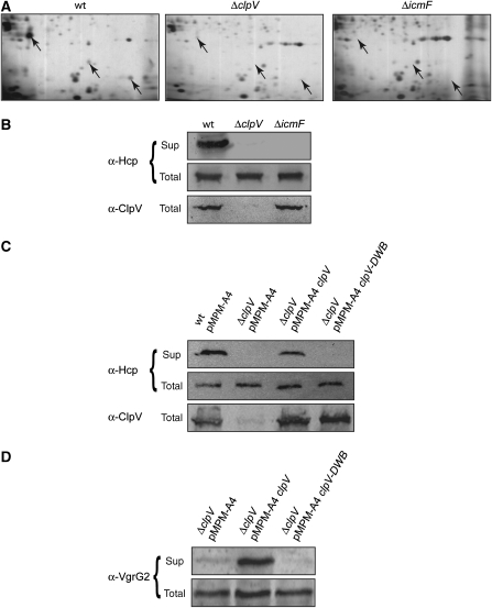
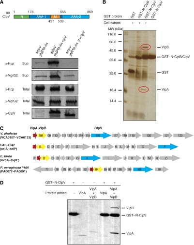
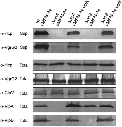
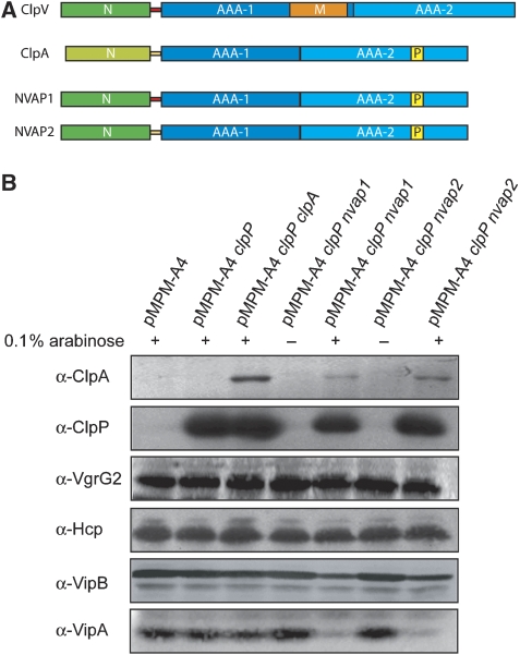
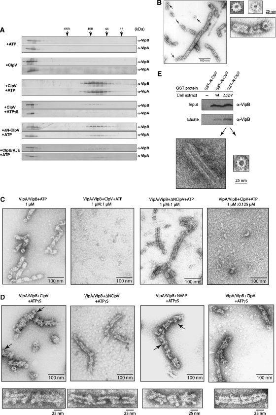
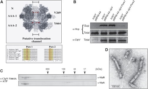
The recently identified type VI secretion systems (T6SS) have a crucial function in the virulence of various proteobacteria, including the human pathogen Vibrio cholerae. T6SS are encoded by a conserved gene cluster comprising approximately 15 open reading frames, mediating the appearance of Hcp and VgrG proteins in cell culture supernatants. Here, we analysed the function of the V. cholerae T6SS member ClpV, a specialized AAA+ protein. ClpV is crucial for a functional T6SS and interacts through its N-terminal domain with the VipA/VipB complex that is composed of two conserved and essential members of T6SS. Transferring ClpV substrate specificity to a distinct AAA+ protein involved in proteolysis caused degradation of VipA but not Hcp or VgrG2, suggesting that VipA rather than Hcp/VgrG2 functions as a primary ClpV substrate. Strikingly, VipA/VipB form tubular, cogwheel-like structures that are converted by a threading activity of ClpV into small complexes. ClpV-mediated remodelling of VipA/VipB tubules represents a crucial step in T6S, illuminating an unexpected role of an ATPase component in protein secretion.


Figures

Figure 1
Figure 1
ClpV is an essential energizing component of T6SS. (A) Culture supernatants of V. cholerae V52 wild-type (V.c. wt) and V. cholerae V52 ΔclpV and ΔicmF mutant cells were separated by 2D gel electrophoresis followed by silver staining. Protein spots that were specifically present in V.c. wt but missing in V.c. ΔclpV and V.c. ΔicmF mutants (arrows) were identified by mass spectrometry as full-length Hcp or Hcp degradation products. (B) The deficiencies of V.c. ΔclpV and V.c. ΔicmF mutants in Hcp secretion were verified by immunoblot analysis using Hcp-specific antibodies. Sup: culture supernatant; total: total cell extract. (C, D) Complementation of the V.c. ΔclpV secretion defect by plasmid-encoded clpV and clpV-DWB using Hcp- or VgrG2-specific antibodies. ClpV levels were monitored by immunoblot analysis using ClpV-specific antibodies.
Figure 2
Figure 2
The ClpV N-domain is essential for ClpV activity and mediates binding to the VipA/VipB complex. (A) Upper part: schematic domain organization of V. cholerae ClpV. N: N-terminal domain; M: middle domain. Domain boundaries are indicated (amino-acid (aa) numbering). Lower part: complementation analysis of the V.c. ΔclpV secretion defect by plasmid-encoded clpV and its deletion variant lacking the N-terminal domain (ΔN-clpV) using Hcp- and VgrG2-specific antibodies. ClpV levels were monitored by immunoblot analysis using ClpV-specific antibodies. Sup: culture supernatant; total: total cell extract. (B) Purified GST–N-ClpV was coupled to glutathione beads and incubated with soluble cell extracts of V. cholerae V52 ΔclpV. Cell extract incubations with either GST or GST–N-ClpB served as a control. Proteins that were specifically co-purified with GST–N-ClpV (red circles) were identified by mass spectrometry as VCA0107 and VCA0108 and were termed VipA (ClpV-interacting protein A) and VipB, respectively. The positioning of GST, GST–N-ClpV and GST–N-ClpB is indicated. A protein standard is given. (C) Schematic representation of the T6SS encoding gene clusters of V. cholerae, enteroaggregative Escherichia coli (EAEC 42), Edwardsiella tarda and Pseudomonas aeruginosa. VipA, VipB and ClpV encoding genes are coloured in red, yellow and blue, respectively. (D) Purified GST–N-ClpV was coupled to glutathione beads and incubated with purified VipA or VipA/VipB complex. Incubation of VipA or VipA/VipB with empty glutathione beads (−GST–N-ClpV) served as a control. The specific co-sedimentation of VipA/VipB with GST–N-ClpV demonstrates complex formation.
Figure 3
Figure 3
VipA and VipB are crucial for T6SS. Analysis of Hcp and VgrG2 secretion in V. cholerae V52 wild-type (wt) and V. cholerae V52 ΔvipA or ΔvipB mutant cells harbouring either a vector control (pMPM-A4) or plasmid-encoded vipA or vipB. Hcp and VgrG2 secretion was monitored by immunoblot analysis using specific antibodies. VipA and VipB levels were determined using VipA- and VipB-specific antibodies. Sup: culture supernatant; total: total cell extract.
Figure 4
Figure 4
Transplanting the ClpV N-domain to the ClpA/ClpP proteolytic machinery results in VipA degradation. (A) Schematic domain organization of V. cholerae ClpV, E. coli ClpA and the hybrid NVAP1/2 (ClpV/ClpA) fusion proteins. The flexible linker sequences, connecting the N-terminal domain and the AAA-1 domain of ClpV or ClpA are coloured in red and yellow, respectively. The P-loop motif (P), which is crucial for the interaction of ClpA with the peptidase ClpP, is indicated. N: N-terminal domain; M: middle domain. (B) V. cholerae V52 ΔclpV mutant cells harbouring the indicated plasmids were grown in the absence or presence of 0.1% (w/v) arabinose as indicated. Total levels of ClpA, ClpP, VgrG, Hcp, VipA and VipB were determined by immunoblot analysis using specific antibodies. A full-colour version of this figure is available at The EMBO Journal Online.
Figure 5
Figure 5
ClpV converts cogwheel-like VipA/VipB tubules to small complexes. (A) VipA/VipB complexes were incubated in the presence of the indicated components for 30 min at 30°C. Complex integrity was monitored by size-exclusion chromatography and eluted fractions were analysed by immunoblotting using VipA- and VipB-specific antibodies. Elution positions of protein standards are indicated by arrows. (B) Morphology of VipA/VipB complexes monitored by electron microscopy. Arrows indicate cogwheel-like structures. Respective scale bars are given. (C) VipA/VipB complexes were incubated without or with ClpV or ΔN-ClpV (+ATP) for 30 min at 30°C and analysed by electron microscopy. Respective scale bars are given. (D) VipA/VipB complexes were incubated with ClpV, ΔN-ClpV, NVAP1 and ClpA (each+ATPγS) and analysed by electron microscopy. Arrows indicate dot-like structures that are associated with VipA/VipB tubules and exhibiting a central hole. Respective scale bars are given. (E) Purified GST–N-ClpV was coupled to glutathione beads and incubated with soluble cell extracts of V. cholerae V52 wild type and ΔclpV. Bound proteins were eluted by the addition of glutathione and analysed by immunoblot analysis using VipB-specific antibodies and by electron microscopy. Equal amounts of VipB present in the used cell extracts were confirmed by western blot (input control).
Figure 6
Figure 6
A conserved pore-located residue of ClpV is crucial for T6S and remodelling of the VipA/VipB complex. (A) Upper part: surface representation of a ClpV hexamer model. Two monomers were removed from the hexamer to show the inside of the putative translocation channel. The positioning of N-terminal domains (N) and ATPase domains (AAA-1 and AAA-2) are indicated. Pore-located Val269 and Tyr664 are shown as CPK models and are coloured in red. Lower part: multiple sequence alignment of polypeptide segments located at the central pore entrance of the first or second AAA domain (pore 1 and pore 2, respectively) of various ClpV proteins. The protein sequence of the pore regions of V. cholerae ClpV was aligned with numerous other ClpV proteins from Salmonella typhimurium, Yersinia pestis, Erwinia carotowora and Rhodobacter sphaeroides. V. cholerae ClpA and ClpB are included as a reference. Conserved, pore-located residues that have been implicated in ClpA- or ClpB-mediated substrate threading are highlighted (red rectangle). (B) Complementation analysis of the V.c. ΔclpV secretion defect by plasmid-encoded clpV and pore mutant derivatives using Hcp-specific antibodies. ClpV levels were monitored by immunoblot analysis using ClpV-specific antibodies. Sup: culture supernatant, total: total cell extract. The ClpV-Y664A pore mutant does not efficiently dissociate VipA/VipB complexes as analysed by size-exclusion chromatography (C) and electron microscopy (D). A scale bar is indicated.

Comment in

References

    1. Akeda Y, Galan JE (2005) Chaperone release and unfolding of substrates in type III secretion. Nature 437: 911–915 - PubMed
    1. Bladergroen MR, Badelt K, Spaink HP (2003) Infection-blocking genes of a symbiotic Rhizobium leguminosarum strain that are involved in temperature-dependent protein secretion. Mol Plant Microbe Interact 16: 53–64 - PubMed
    1. Cascales E (2008) The type VI secretion toolkit. EMBO Rep 9: 735–741 - PMC - PubMed
    1. Das S, Chaudhuri K (2003) Identification of a unique IAHP (IcmF associated homologous proteins) cluster in Vibrio cholerae and other proteobacteria through in silico analysis. In Silico Biol 3: 287–300 - PubMed
    1. de Bruin OM, Ludu JS, Nano FE (2007) The Francisella pathogenicity island protein IglA localizes to the bacterial cytoplasm and is needed for intracellular growth. BMC Microbiol 7: 1. - PMC - PubMed

Publication types