. 2009 Jan;5(1):e1000259.
doi: 10.1371/journal.ppat.1000259. Epub 2009 Jan 9.

A novel co-crystal structure affords the design of gain-of-function lentiviral integrase mutants in the presence of modified PSIP1/LEDGF/p75

Affiliations

A novel co-crystal structure affords the design of gain-of-function lentiviral integrase mutants in the presence of modified PSIP1/LEDGF/p75

Stephen Hare et al. PLoS Pathog. 2009 Jan.

Abstract

Lens epithelium derived growth factor (LEDGF), also known as PC4 and SFRS1 interacting protein 1 (PSIP1) and transcriptional co-activator p75, is the cellular binding partner of lentiviral integrase (IN) proteins. LEDGF accounts for the characteristic propensity of Lentivirus to integrate within active transcription units and is required for efficient viral replication. We now present a crystal structure containing the N-terminal and catalytic core domains (NTD and CCD) of HIV-2 IN in complex with the IN binding domain (IBD) of LEDGF. The structure extends the known IN-LEDGF interface, elucidating primarily charge-charge interactions between the NTD of IN and the IBD. A constellation of acidic residues on the NTD is characteristic of lentiviral INs, and mutations of the positively charged residues on the IBD severely affect interaction with all lentiviral INs tested. We show that the novel NTD-IBD contacts are critical for stimulation of concerted lentiviral DNA integration by LEDGF in vitro and for its function during the early steps of HIV-1 replication. Furthermore, the new structural details enabled us to engineer a mutant of HIV-1 IN that primarily functions only when presented with a complementary LEDGF mutant. These findings provide structural basis for the high affinity lentiviral IN-LEDGF interaction and pave the way for development of LEDGF-based targeting technologies for gene therapy.


Conflict of interest statement

The authors have declared that no competing interests exist.

Figures

Figure 1
Figure 1. The HIV-2 INNTD+CCD:LEDGFIBD Structure and Multimeric Assemblies Found in the Crystal.
(A) The IN2LEDGF substructure, containing a dimer of INNTD+CCD and a single molecule of LEDGFIBD, the basic building unit of both crystal forms. IN chains are colored pale green and cyan, and LEDGF is pink. Zinc atoms are shown as gray spheres. Residues involved in IN–LEDGF interfaces and discussed in the text are shown as sticks and indicated with arrowheads. (B) The closed trimer of IN2LEDGF substructures. Colors and labels as in (A). (C) Stereo view of the higher order assembly involving four IN2LEDGF trimers representing the entire ASU of crystal form II. Colors as in (A).
Figure 2
Figure 2. Details of the NTD–IBD Interface.
(A) The acidic IN residues and basic LEDGF residues are shown as sticks. The 2Fo-Fc electron density map, contoured at 1σ, is shown in pale blue. (B) Partial amino acid sequence alignment including residues 1–17 of human (HIV-1 and HIV-2) and non-primate (BIV, EIAV, FIV, and MVV) lentiviral INs. Conserved residues are shown in bold; His-12 and His-16 of the invariant HHCC motif are highlighted in yellow. Acidic residues known (for HIV-1 and HIV-2) or proposed to interact with the positively charged face of the IBD are orange.
Figure 3
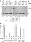
Figure 3. Functional Importance of the NTD–IBD Charge–Charge Interface for the Lentiviral IN–LEDGF Interaction.
(A) Pull down experiments. Non-tagged WT, D366N, AAA, ESE, EEE, K360E, or K392E LEDGF proteins were incubated with C-terminally His6-tagged forms of HIV-1 IN mutant H12N, WT HIV-1 IN, HIV-2 IN, BIV IN, MVV IN, or EIAV IN (as indicated) in the presence of Ni-NTA agarose. Bound proteins were separated in 11% sodium dodecyl sulfate polyacrylamide gels and visualized with Coomassie Blue. Input quantities of LEDGF proteins are shown below the main gel. (B) Yeast two-hybrid analyses. The IN–LEDGF interaction assay , is based on transactivation of the β-galactosidase gene in S. cerevisiae indicator cells Y187. Relative β-galactosidase activities produced by yeast co-transformed with the indicated IN and LEDGFIBD mutants are shown on a log scale, with 100% corresponding to the WT bait/WT prey condition. The background of the assay is defined by WT IN in the presence of empty prey vector and is shown as a gray line. Each bar represents a mean value; standard deviations were calculated based on results of quadruplicate measurements. Values exceeding the background are shown atop the bars.
Figure 4
Figure 4. Effects of Mutations within the NTD–IBD Interface on LEDGF–Dependent Strand Transfer Activities of Lentiviral INs.
(A) Schematic of the reactions mediated by IN in the presence of circular DNA target: concerted integration results in a linear product while half-site reactions produce branched molecules (circular half-site and donor-donor). (B) HIV-1 IN activity under established conditions that favor half-site integration . WT, KK, or KKK HIV-1 IN was incubated with 10 nM 500-bp donor DNA and supercoiled target DNA in the absence (lane 2) or presence (lanes 3–11) of WT, D366N, or EEE LEDGF. The products separated in an agarose gel were detected with ethidium bromide. Lane 1 contained a mock sample, with both IN and LEDGF omitted. (C) EIAV IN reactions. EIAV IN was incubated with 10 nM 225-bp donor DNA and supercoiled target DNA in the absence (lane 2) or presence (lanes 3 and 4) of WT or EEE LEDGF, as indicated. Lane 1 contained a mock sample without IN and LEDGF; lane 5 contained IN and WT LEDGF without donor DNA; lane 6 contained 10 ng of singly-nicked pGEM-9Zf(-) as a migration standard for the open circular (o.c.) form of target DNA. (D) Enhanced concerted HIV-1 integration assay using an elevated input of donor DNA (for details, see Text S1 and Figure S4). WT, KK, or KKK HIV-1 IN was incubated with 0.5 µM pre-processed 32-bp donor and supercoiled target DNA in the presence of WT, D366N, AAA, ESE, EEE, K360E, or K392E LEDGF. Migration positions of various DNA species (size standards, supercoiled [s.c.] and o.c. target, donor, and reaction products) are indicated.
Figure 5
Figure 5. LEDGF Residues Lys-401, Arg-404, and Arg-405 Play a Crucial Role in HIV-1 Infection.
(A) Relative infectivity of VSV G-pseudotyped WT HIV-Luc virus on Ledgf-null E2 MEFs transfected with human WT, AAA, EAE, EEE, E4, or D366N LEDGF expression vectors, as indicated. Infectivity of the virus in the presence of WT LEDGF was set to 100%; mean values and standard deviations combine data of two independent sets of transfections (each with infections performed in duplicate). (B) Infectivity of HIV-Luc bearing KK mutations in IN on E2 MEFs transfected with WT, EAE, or EEE LEDGF. Infectivity is expressed as a percentage of WT virus on the cells transfected with WT LEDGF. Western blots above the graphs show expression levels of LEDGF proteins and actin loading control.

References

    1. Lewinski MK, Bushman FD. Retroviral DNA integration–mechanism and consequences. Adv Genet. 2005;55:147–181. - PubMed
    1. Craigie R. Retroviral DNA Integration. In: Craig NL, Craigie R, Gellert M, Lambowitz AM, editors. Mobile DNA II. Washington DC: ASM Press; 2002. pp. 613–630.
    1. Bushman FD, Engelman A, Palmer I, Wingfield P, Craigie R. Domains of the integrase protein of human immunodeficiency virus type 1 responsible for polynucleotidyl transfer and zinc binding. Proc Natl Acad Sci USA. 1993;90:3428–3432. - PMC - PubMed
    1. Engelman A, Bushman FD, Craigie R. Identification of discrete functional domains of HIV-1 integrase and their organization within an active multimeric complex. EMBO J. 1993;12:3269–3275. - PMC - PubMed
    1. Engelman A, Craigie R. Identification of conserved amino acid residues critical for human immunodeficiency virus type 1 integrase function in vitro. J Virol. 1992;66:6361–6369. - PMC - PubMed

Publication types

Substances