

Disease modeling for Ebola and Marburg viruses
- PMID: 19132113
- PMCID: PMC2615158
- DOI: 10.1242/dmm.000471
Disease modeling for Ebola and Marburg viruses
Abstract
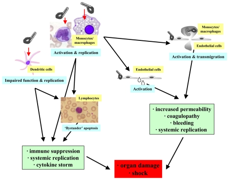
The filoviruses Ebola and Marburg are zoonotic agents that are classified as both biosafety level 4 and category A list pathogens. These viruses are pathogenic in humans and cause isolated infections or epidemics of viral hemorrhagic fever, mainly in Central Africa. Their natural reservoir has not been definitely identified, but certain species of African bat have been associated with Ebola and Marburg infections. Currently, there are no licensed options available for either treatment or prophylaxis. Different animal models have been developed for filoviruses including mouse, guinea pig and nonhuman primates. The 'gold standard' animal models for pathogenesis, treatment and vaccine studies are rhesus and cynomolgus macaques. This article provides a brief overview of the clinical picture and the pathology/pathogenesis of human filovirus infections. The current animal model options are discussed and compared with regard to their value in different applications. In general, the small animal models, in particular the mouse, are the most feasible for high biocontainment facilities and they offer the most options for research owing to the greater availability of immunologic and genetic tools. However, their mimicry of the human diseases as well as their predictive value for therapeutic efficacy in primates is limited, thereby making them, at best, valuable initial screening tools for pathophysiology, treatment and vaccine studies.
Figures
References
-
- Aleksandrowicz P., Wolf K., Falzarano D., Feldmann H., Seebach J., Schnittler H. (2008). Viral haemorrhagic fever and vascular alterations. Hamostaseologie 28, 77–84 - PubMed
-
- Borisevich I. V., Mikhailov V. V., Krasnianskii V. P., Gradoboev V. N., Lebedinskaia E. V., Potryvaeva N. V., Timan’kova G. D. (1995). Development and study of the properties of immunoglobulin against Ebola fever. Vopr. Virusol. 40, 270–273 - PubMed
-
- Bowen E. T., Platt G. S., Simpson D. I., McArdell L. B., Raymond R. T. (1978). Ebola haemorrhagic fever: experimental infection of monkeys. Trans. R. Soc. Trop. Med. Hyg. 72, 188–191 - PubMed
-
- Bowen E. T., Platt G. S., Lloyd G., Raymond R. T., Simpson D. I. (1980). A comparative study of strains of Ebola virus isolated from southern Sudan and northern Zaire in 1976. J. Med. Virol. 6, 129–138 - PubMed
-
- Bradfute S. B., Braun D. R., Shamblin J. D., Geisbert J. B., Paragas J., Garrison A., Hensley L. E., Geisbert T. W. (2007). Lymphocyte death in a mouse model of Ebola virus infection. J. Infect. Dis. 196 Suppl. 2, S296–S304 - PubMed
MeSH terms
LinkOut - more resources
Full Text Sources
Other Literature Sources
Medical
