. 2009 Feb;9(3):671-85.
doi: 10.1002/pmic.200800441.

Proteomic profiling of antisense-induced exon skipping reveals reversal of pathobiochemical abnormalities in dystrophic mdx diaphragm

Affiliations

Proteomic profiling of antisense-induced exon skipping reveals reversal of pathobiochemical abnormalities in dystrophic mdx diaphragm

Philip Doran et al. Proteomics. 2009 Feb.

Abstract

The disintegration of the dystrophin-glycoprotein complex represents the initial pathobiochemical insult in Duchenne muscular dystrophy. However, secondary changes in signalling, energy metabolism and ion homeostasis are probably the main factors that eventually cause progressive muscle wasting. Thus, for the proper evaluation of novel therapeutic approaches, it is essential to analyse the reversal of both primary and secondary abnormalities in treated muscles. Antisense oligomer-mediated exon skipping promises functional restoration of the primary deficiency in dystrophin. In this study, an established phosphorodiamidate morpholino oligomer coupled to a cell-penetrating peptide was employed for the specific removal of exon 23 in the mutated mouse dystrophin gene transcript. Using DIGE analysis, we could show the reversal of secondary pathobiochemical abnormalities in the dystrophic diaphragm following exon-23 skipping. In analogy to the restoration of dystrophin, beta-dystroglycan and neuronal nitric oxide synthase, the muscular dystrophy-associated differential expression of calsequestrin, adenylate kinase, aldolase, mitochondrial creatine kinase and cvHsp was reversed in treated muscle fibres. Hence, the re-establishment of Dp427 coded by the transcript missing exon 23 has counter-acted dystrophic alterations in Ca2+-handling, nucleotide metabolism, bioenergetic pathways and cellular stress response. This clearly establishes the exon-skipping approach as a realistic treatment strategy for diminishing diverse downstream alterations in dystrophinopathy.


Figures
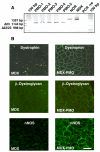
Figure 1
Figure 1. Polymerase chain reaction and immunofluorescence analysis of dystrophin in antisense oligomer-treated MDX diaphragm
Shown is the RT-PCR analysis of normal, MDX and PMO-treated diaphragm tissue. The product of 1357 bp represents the full-length dystrophin transcript, whereas the products of 1144 bp and 998 bp represent transcripts excluding exon 23, and exons 22 and 23, respectively (A). PMO-1 to PMO-5 represents preparations from 5 individual MDX mice treated with antisense oligonucleotides. The outer lanes contain a 100 bp ladder. In panel (B) is shown the restoration of surface expression of dystrophin, β-dystroglycan, and the neuronal nNOS isoform of nitric oxide synthase in PMO-treated MDX tissue as compared to untreated MDX diaphragm muscle. Immunofluorescence microscopy was performed with 6μm unfixed cryosections. Bar equals 50μm.
Figure 2
Figure 2. Overview of DIGE analysis of antisense oligonucleotide-induced exon skipping in dystrophic MDX diaphragm
Shown is the composition of a set of 6 analytical DIGE gels (A-F). To conform to best experimental practice, a randomized labeling protocol was performed. Four sample replicates from each of the three experimental populations (NOR, normal diaphragm; MDX; dystrophic diaphragm; PMO, phosphorodiamidate morpholino oligomer-treated dystrophic diaphragm) were either labeled with Cy3 or Cy5 minimal dyes. Samples were both evenly distributed between CyDye Fluors and between analytical gels. A pool of all samples was prepared and labelled with Cy2 to be used as a standard on all gels to aid image matching and cross gel statistical analysis.
Figure 3
Figure 3. Two-dimensional gel electrophoretic analysis of dystrophic diaphragm following phosphorodiamidate morpholino oligomer-induced exon skipping
Shown is a Cy2-labelled master gel of the total soluble protein complement from normal diaphragm versus dystrophic diaphragm versus phosphorodiamidate morpholino oligomer-treated dystrophic diaphragm muscle. Muscle proteins with a differential expression level are marked by circles and are numbered 1 to 20. See Table 1 for a detailed listing of skeletal muscle proteins with a changed abundance in dystrophic or treated fibres. The pH-values of the first dimension gel system and molecular mass standards (in kDa) of the second dimension are indicated on the top and on the left of the panels, respectively.
Figure 4
Figure 4. Biochemical status of muscular dystrophy-associated markers in antisense oligomer-treated MDX diaphragm
Shown are expanded views of two-dimensional gels of the CyDye-labelled samples from normal diaphragm versus dystrophic MDX diaphragm versus phosphorodiamidate morpholino oligomer-treated dystrophic diaphragm muscle (MDX-PMO), as well as the comparative graphic representation of protein spots representing adenylate kinase (AK), isocitrate dehydrogenase (ICDH), aldolase (ALD), and the small heat shock protein cvHsp.
Figure 5
Figure 5. Two-dimensional gel electrophoretic analysis of dystrophic versus phosphorodiamidate morpholino oligomer-treated diaphragm
Shown is a Cy2-labelled master gel of the total soluble protein complement from dystrophic diaphragm versus phosphorodiamidate morpholino oligomer treated dystrophic diaphragm muscle (A). Muscle proteins with a differential expression level are marked by circles and are numbered 1 to 15. See Table 2 for a detailed listing of skeletal muscle proteins with a changed abundance in dystrophic or treated fibres. The pH-values of the first dimension gel system and molecular mass standards (in kDa) of the second dimension are indicated on the top and on the left of the panels, respectively. In panels (B) to (D) are shown representative images of two-dimensional gels with Cy2-labelled pooled standard, Cy3-labelled dystrophic MDX preparations, and Cy5-labelled phosphorodiamidate morpholino oligomer-treated dystrophic samples (MDX-PMO), respectively.
Figure 6
Figure 6. Proteomic profiling of dystrophic versus antisense oligomer-treated MDX diaphragm
Shown are expanded views of two-dimensional gels of the CyDye-labelled samples from dystrophic MDX diaphragm versus phosphorodiamidate morpholino oligomer-treated dystrophic diaphragm muscle (MDX-PMO), as well as the comparative graphic representation of protein spots representing the small heat shock protein cvHsp, adenylate kinase (AK) and the mitochondrial creatine kinase (mCK).
Figure 7
Figure 7. Two-dimensional immunoblot analysis of muscular dystrophy-associated markers in antisense oligomer-treated MDX diaphragm
Shown is an expanded view of immuno-decorated 2-D spots representing the dystrophin-associated surface glycoprotein β-dystroglycan (β-DG), the sarcoplasmic reticulum Ca2+-binding protein calsequestrin (CSQ), the small heat shock protein cvHsp, carbonic anhydrase isoform CA3, adenylate kinase isoform AK1 and isocitrate dehydrogenase (ICDH) in normal diaphragm versus dystrophic MDX diaphragm versus phosphorodiamidate morpholino oligomer-treated dystrophic diaphragm muscle (MDX-PMO). The position of immuno-decorated spots is marked by arrowheads.

References

    1. Emery AE. The muscular dystrophies. Lancet. 2002;359:687–695. - PubMed
    1. Ahn AH, Kunkel LM. The structural and functional diversity of dystrophin. Nat. Genet. 1993;3:283–291. - PubMed
    1. Chamberlain JS. Gene therapy of muscular dystrophy. Hum. Mol. Genet. 2002;11:2355–2362. - PubMed
    1. Van Deutekom JC, van Ommen GJ. Advances in Duchenne muscular dystrophy gene therapy. Nat. Rev. Genet. 2003;4:774–783. - PubMed
    1. Bogdanovich S, Krag TOB, Barton ER, Morris LD, et al. Functional improvement of dystrophic muscle by myostatin blockade. Nature. 2002;420:418–421. - PubMed

Publication types

MeSH terms