. 2009 Apr 15;419(2):317-28.
doi: 10.1042/BJ20081364.

The ABC transporter MsbA interacts with lipid A and amphipathic drugs at different sites

Affiliations

The ABC transporter MsbA interacts with lipid A and amphipathic drugs at different sites

Alena Siarheyeva et al. Biochem J. .

Abstract

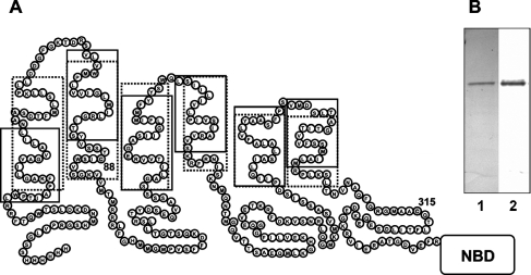
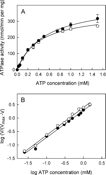
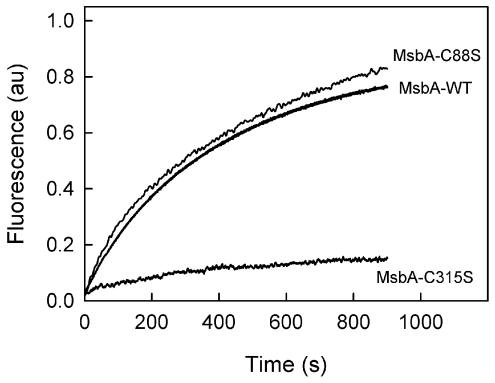
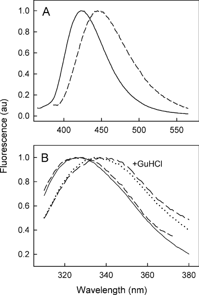
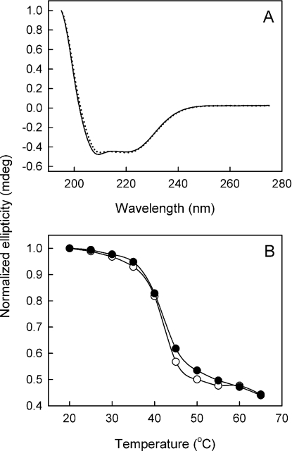
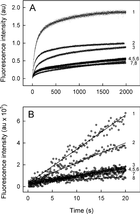
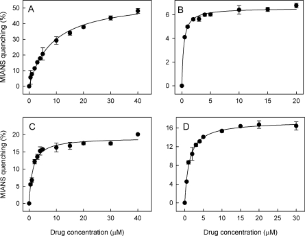
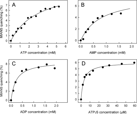
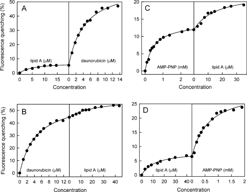
MsbA is an essential ABC (ATP-binding cassette) transporter involved in lipid A transport across the cytoplasmic membrane of Gram-negative bacteria. The protein has also been linked to efflux of amphipathic drugs. Purified wild-type MsbA was labelled stoichiometrically with the fluorescent probe MIANS [2-(4'-maleimidylanilino)naphthalene-6-sulfonic acid] on C315, which is located within the intracellular domain connecting transmembrane helix 6 and the nucleotide-binding domain. MsbA-MIANS displayed high ATPase activity, and its folding and stability were unchanged. The initial rate of MsbA labelling by MIANS was reduced in the presence of amphipathic drugs, suggesting that binding of these compounds alters the protein conformation. The fluorescence of MsbA-MIANS was saturably quenched by nucleotides, lipid A and various drugs, and estimates of the Kd values for binding fell in the range of 0.35-10 microM. Lipid A and daunorubicin were able to bind to MsbA-MIANS simultaneously, implying that they occupy different binding sites. The effects of nucleotide and lipid A/daunorubicin binding were additive, and binding was not ordered. The Kd of MsbA for binding lipid A was substantially decreased when the daunorubicin binding site was occupied first, and prior binding of nucleotide also modulated lipid A binding affinity. These results indicate that MsbA contains two substrate-binding sites that communicate with both the nucleotide-binding domain and with each other. One is a high affinity binding site for the physiological substrate, lipid A, and the other site interacts with drugs with comparable affinity. Thus MsbA may function as both a lipid flippase and a multidrug transporter.


Figures

Figure 1
Figure 1. Topology and purification of MsbA
(A) Topological model of the MsbA protein. Each monomer contains 6 membrane-spanning helices forming a TM domain, with the NBD at the C-terminal end. Amino acid residues are indicated inside the circles; endogenous cysteine residues are numbered. The dotted box represents the TM domain boundaries of MsbA determined by EPR spectroscopy [21]. The X-ray crystal structure of Salmonella typhimurium MsbA (PDB: 3b60 [15]) suggests the following membrane-spanning domain boundaries: 1(27–52), 2(62–87), 3(142–163), 4(165–182), 5(248–271) and 6(283–305). The solid box represents the predicted TM domain boundaries (http://www.expasy.org/uniprot/P60752). (B) Purification of MsbA from cytoplasmic membrane vesicles of E. coli overexpressing the protein. Lane 1, SDS/PAGE analysis of purified MsbA (1 μg) in 0.05% (w/v) DM, stained with Coomassie Blue; lane 2, Western blot of purified MsbA using an anti-His6 antibody.
Figure 2
Figure 2. Catalytic activity of MsbA and MsbA–MIANS
(A) The ATPase activity at 37 °C as a function of ATP concentration for MsbA–MIANS (○) and unlabelled MsbA (●). Data points are the means±S.D. (n=3). In both cases, purified MsbA was in a buffer containing 0.05% (w/v) DM. (B) Hill plot of the ATPase activity of wild-type MsbA (●) and MsbA–MIANS (○) as a function of ATP concentration; MsbA Hill coefficient, n=1.03±0.03, MsbA–MIANS Hill coefficient, n=1.01±0.03.
Figure 3
Figure 3. Reaction of wild-type and cysteine mutant MsbA proteins with MIANS
Time-dependent reaction of wild-type MsbA (MsbA-WT) and the site-directed mutants MsbA-C88S and MsbA-C316S with MIANS. MIANS labelling of MsbA (100 μg/ml) was carried out in buffer containing 0.05% (w/v) DM. Labelling was initiated by addition of 10 μM MIANS, and fluorescence emission was recorded continuously at 420 nm (λex=322 nm).
Figure 4
Figure 4. Fluorescence emission spectra of MsbA and MsbA–MIANS
(A) Fluorescence emission spectra of MIANS (λex=322 nm) covalently linked to MsbA (continuous line) and to the soluble compound DTE (broken line). (B) Intrinsic Trp fluorescence emission spectra (λex=290 nm) of MsbA–MIANS (short-dashed line and long-dashed line) and unlabelled MsbA (continuous line and dotted line) before and after, respectively, treatment with 6 M guanidine hydrochloride (GuHCl).
Figure 5
Figure 5. CD spectroscopy of MsbA and MsbA–MIANS
(A) CD spectra of unlabelled MsbA (continuous line) and MsbA–MIANS (dotted line) at a concentration of 0.35 mg/ml in buffer with 0.01% (w/v) DM. (B) Thermal unfolding of MsbA and MsbA–MIANS monitored by CD spectroscopy. CD measurements were carried out on purified unlabelled MsbA (●) and MsbA–MIANS (○) at a concentration of 0.35 mg/ml in buffer with 0.01% (w/v) DM. Molar ellipticity was recorded at 222 nm, which reports on α-helical unfolding.
Figure 6
Figure 6. Rate of reaction of MsbA with MIANS in the presence of drugs and lipid A
MIANS labelling of MsbA (70 μg/ml) was carried out in buffer containing 0.05% (w/v) DM. MsbA was preincubated for 15 min at 23 °C with various drugs and lipid A at concentrations (see Table 1) corresponding to ∼5-fold higher than their Kd for binding (see Table 2). (A) Labelling was initiated by adding MIANS (20 μM) and fluorescence emission intensity fluorescence emission was recorded continuously (0.2 s intervals) at 420 nm (λex=322 nm), for times up to 2000 s. (B) Initial rates of labelling were observed over the first 20 s after addition of MIANS. 1, MsbA in the absence of drug; 2, 6 μM vinblastine; 3, 40 μM quercetin; 4, 6 μM verapamil; 5, 2 μM valinomycin; 6, 30 μM lipid A; 7, 4 μM propafenone GP12; and 8, 25 μM daunorubicin.
Figure 7
Figure 7. Quenching of MsbA–MIANS fluorescence by drugs
Increasing concentrations of various drugs were added to 100 μg/ml MsbA–MIANS in 0.05% (w/v) DM buffer at 23 °C. The fluorescence emission was monitored at 420 nm (λex=322 nm). The percent quenching of MsbA–MIANS fluorescence (ΔF/F0×100) was calculated relative to the fluorescence in the absence of drugs. The continuous line represents the best computer-generated fit of the data points to an equation describing interaction with a single type of binding site, and were used to estimate the Kd of binding. (A) quercetin, (B) valinomycin, (C) verapamil and (D) vinblastine. Data points are the means±S.D. (n=3).
Figure 8
Figure 8. Quenching of MsbA–MIANS fluorescence by nucleotides
Increasing concentrations of nucleotides were added to 100 μg/ml MsbA–MIANS in 0.05% (w/v) DM buffer at 23 °C (with exception of ATP, which was added to MsbA at 10 °C to prevent ATP hydrolysis). The fluorescence emission was monitored at 420 nm (λex=322 nm). The percent quenching of MsbA–MIANS fluorescence (ΔF/F0×100) was calculated relative to the fluorescence in the absence of nucleotides. The continuous line represents the best computer-generated fit of the data points to an equation describing interaction with a single type of binding site, and were used to estimate the Kd of binding. (A) ATP, (B) AMP, (C) ADP and (D) ATP[S] (ATPγS). Data points are the means±S.D. (n=3).
Figure 9
Figure 9. Sequential binding of lipid A, daunorubicin and nucleotides to MsbA
Dual sequential titrations of MsbA–MIANS in 0.05% DM buffer with lipid A, daunorubicin and p[NH]ppA (AMP-PNP) were performed at 23 °C, with fluorescence emission monitored at 420 nm (λex=322 nm). Titration with (A) lipid A and then daunorubicin, (B) daunorubicin and then lipid A, (C) p[NH]ppA and then lipid A, (D) lipid A and then p[NH]ppA. The continuous lines represent the best computer-generated fit of the data points to an equation describing interaction with a single type of binding site, and were used to estimate the Kd of binding. Data points are the means of three experiments.

References

    1. Dassa E., Bouige P. The ABC of ABCs: a phylogenetic and functional classification of ABC systems in living organisms. Res. Microbiol. 2001;152:211–229. - PubMed
    1. Davidson A. L., Chen J. ATP-binding cassette transporters in bacteria. Annu. Rev. Biochem. 2004;73:241–268. - PubMed
    1. Higgins C. F. ABC transporters: physiology, structure and mechanism – an overview. Res. Microbiol. 2001;152:205–210. - PubMed
    1. Doerrler W. T., Reedy M. C., Raetz C. R. An Escherichia coli mutant defective in lipid export. J. Biol. Chem. 2001;276:11461–11464. - PubMed
    1. Doerrler W. T., Gibbons H. S., Raetz C. R. MsbA-dependent translocation of lipids across the inner membrane of Escherichia coli. J. Biol. Chem. 2004;279:45102–45109. - PubMed

Publication types

MeSH terms

LinkOut - more resources