Review
. 2009 Feb 27;61(2):158-71.
doi: 10.1016/j.addr.2008.11.002. Epub 2008 Dec 13.

Mucus-penetrating nanoparticles for drug and gene delivery to mucosal tissues

Affiliations
Review

Mucus-penetrating nanoparticles for drug and gene delivery to mucosal tissues

Samuel K Lai et al. Adv Drug Deliv Rev. .

Abstract

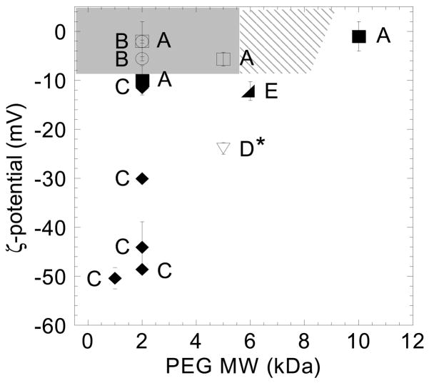
Mucus is a viscoelastic and adhesive gel that protects the lung airways, gastrointestinal (GI) tract, vagina, eye and other mucosal surfaces. Most foreign particulates, including conventional particle-based drug delivery systems, are efficiently trapped in human mucus layers by steric obstruction and/or adhesion. Trapped particles are typically removed from the mucosal tissue within seconds to a few hours depending on anatomical location, thereby strongly limiting the duration of sustained drug delivery locally. A number of debilitating diseases could be treated more effectively and with fewer side effects if drugs and genes could be more efficiently delivered to the underlying mucosal tissues in a controlled manner. This review first describes the tenacious mucus barrier properties that have precluded the efficient penetration of therapeutic particles. It then reviews the design and development of new mucus-penetrating particles that may avoid rapid mucus clearance mechanisms, and thereby provide targeted or sustained drug delivery for localized therapies in mucosal tissues.


Figures

Figure 1
Figure 1
Transmission electron micrograph of human cervicovaginal mucus. The interfiber spacing ranges from 10 nm to 200 nm. Individual fibers can be observed with fiber diameters ~10 nm. Scale bar = 200 nm. Figure obtained from [19].
Figure 2
Figure 2
Fluorescently labeled 59 nm polystyrene particles formed thick cables with mucin fibers in human cervicovaginal mucus. (A) Fluorescent image. (B) Phase image. Scale bar = 500 μm. Figure obtained from [19].
Figure 3
Figure 3
Schematic drawing of a Transwell-Snapwell diffusion chamber (A). The donor and acceptor chambers are indicated by I and II, respectively. Mucus or mucin gel is sandwiched in between polycarbonate filters attached to the Snapwell ring (E) located between the two chambers. Figure obtained from [112].
Figure 4
Figure 4
Ensemble-averaged mean square displacement (MSD) as a function of time scale for PLGA-DDAB/DNA and COOH-modified polystyrene nanoparticles in reconstituted pig gastric mucus. Figure obtained from [119].
Figure 5
Figure 5
Agglomerated non-adhesive particles coated with a mucus plug collected immediately following discharge from the proximal jejunum of the perfused rat (magnification: ×60). Figure obtained from [123].
Figure 6
Figure 6
Summary schematic illustrating the fate of mucus-penetrating particles (MPP) and conventional mucoadhesive particles (CP) administered to a mucosal surface. MPP readily penetrate the luminal mucus layer (LML) and enter the underlying adherent mucus layer (AML). In contrast, CP are largely immobilized in the LML. Because MPP can enter the AML and thus are in closer proximity to the cells, cells will be exposed to a greater dose of drug released from MPP compared to drug released from CP. As the LML layer is cleared, CP are removed along with the LML whereas MPP in the AML are retained, leading to prolonged residence time for MPP at the mucosal surface. Thus, at long times, there is almost no drug dosing to cells with CP, whereas MPP, because they are retained longer, will continue to release drugs to cells. Since MPP can penetrate both the LML and AML, a fraction may reach and bind to the underlying epithelia and further improve drug delivery. While this schematic reflects the mucosal physiology of the gastrointestinal and cervicovaginal tracts, the same behavior is expected for the respiratory airways. In the respiratory airways, CP are mostly immobilized in the luminal stirred mucus gel layer, whereas MPP penetrate the mucus gel and enter the underlying periciliary layer. Upon mucociliary clearance, a significant fraction of MPP remains in the periciliary layer, resulting in prolonged retention. This schematic does not depict the glycocalyx adjacent to the epithelial surface, which may contribute an additional steric barrier to cellular entry of MPP.
Figure 7
Figure 7
Normalized diffusion coefficients for proteins and viruses in mucus. If a particle diffuses in mucus as fast as it diffuses in saline, Dmuc/Dpbs = 1. The lines drawn on the graph are the ratios predicted by Amsden’s obstruction-scaling model using a mucin fiber radius of 3.5 nm and mesh fiber spacing of 100 nm (solid line) or 110 nm (dotted line), which accounts for a 10% increase in the mesh fiber spacing due to 20% dilution of the mucus sample. Figure obtained from [19].
Figure 8
Figure 8
Schematic representation of (A) the interdiffusion of polymers upon contact and (B) the interpenetration between tethered chains on a particle surface and mucin fibers of the mucus gel layer. Figure obtained from [175].
Figure 9
Figure 9
Transport of COOH- and PEG-modified polystyrene particles in fresh, human cervicovaginal mucus (CVM). Ensemble-averaged geometric mean square displacements () as a function of time scale for (A) COOH- and (B) PEG-modified polystyrene particles in human CVM. Comparison of average Deff at a time scale of 1 s in water (W) vs. CVM of subfractions of (C) COOH- and (D) PEG-modified particles, from fastest to slowest. Theoretical Deff for same-sized particles in water is shown as W. The dashed black line at <Deff> = 1 × 10⁻⁴ signifies the microscope’s resolution. Data represent ensemble average of three experiments, with n ≥ 120 particles for each experiment. Figure obtained from [17].
Figure 10
Figure 10
Phase diagram correlating muco-inert versus mucoadhesive particle behavior to surface charge and PEG MW for various PEG-coated nanoparticles (~200–500 nm in size) reported in literature. PEG-coated nanoparticles reported to be non-mucoadhesive compared to control (uncoated) particles are indicated by open symbols, and those reported to be mucoadhesive compared to control particles are indicated by filled symbols. The shaded region represents the confirmed range of PEG MW and particle ξ-potential (i.e., PEG surface coverage), and the hatched region an additional predicted range, that provides a muco-inert coating. Letters represent published results from various studies [, , –235]. *Mucoadhesion was not observed based on adhesion to an in vitro mucin-secreting cell line, which is unlikely to produce mucus gels with the complex mesh structure and adhesivity of physiological mucus [234]. Figure obtained from [183].

References

    1. Rosen H, Abribat T. The rise and rise of drug delivery. Nat Rev Drug Discov. 2005;4:381–5. - PubMed
    1. Shmulewitz A, Langer R, Patton J. Convergence in biomedical technology. Nat Biotech. 2006;24:277–277.
    1. Duncan R. The dawning era of polymer therapeutics. Nat Rev Drug Discov. 2003;2:347–60. - PubMed
    1. Allen TM, Cullis PR. Drug delivery systems: entering the mainstream. Science. 2004;303:1818–22. - PubMed
    1. Langer R. Drug delivery and targeting. Nature. 1998;392:5–10. - PubMed

Publication types