Review
. 2009 Feb 1;417(3):639-50.
doi: 10.1042/BJ20080413.

Repair of ionizing radiation-induced DNA double-strand breaks by non-homologous end-joining

Affiliations
Review

Repair of ionizing radiation-induced DNA double-strand breaks by non-homologous end-joining

Brandi L Mahaney et al. Biochem J. .

Abstract

DNA DSBs (double-strand breaks) are considered the most cytotoxic type of DNA lesion. They can be introduced by external sources such as IR (ionizing radiation), by chemotherapeutic drugs such as topoisomerase poisons and by normal biological processes such as V(D)J recombination. If left unrepaired, DSBs can cause cell death. If misrepaired, DSBs may lead to chromosomal translocations and genomic instability. One of the major pathways for the repair of IR-induced DSBs in mammalian cells is NHEJ (non-homologous end-joining). The main proteins required for NHEJ in mammalian cells are the Ku heterodimer (Ku70/80 heterodimer), DNA-PKcs [the catalytic subunit of DNA-PK (DNA-dependent protein kinase)], Artemis, XRCC4 (X-ray-complementing Chinese hamster gene 4), DNA ligase IV and XLF (XRCC4-like factor; also called Cernunnos). Additional proteins, including DNA polymerases mu and lambda, PNK (polynucleotide kinase) and WRN (Werner's Syndrome helicase), may also play a role. In the present review, we will discuss our current understanding of the mechanism of NHEJ in mammalian cells and discuss the roles of DNA-PKcs and DNA-PK-mediated phosphorylation in NHEJ.


Figures

Figure 1
Figure 1. A model for NHEJ
(A) IR induces multiple forms of DNA damage including DSBs that contain non-ligatable end groups such as 3’-phosphate and 3’-phosphoglycolate groups (indicated by ◆). (B) The Ku heterodimer (orange) binds the ends of the DSB, tethering the ends together. Recruitment of Ku to the DSB occurs independently of other known NHEJ or DSB repair proteins, consistent with Ku acting as the cornerstone of the NHEJ pathway. (C) Ku translocates inwards, allowing recruitment of DNA-PKcs (blue) such that it binds the extreme termini of the break (D). Recruitment of DNA-PKcs to the DSB requires Ku but no other NHEJ or DSB repair factors. Two DNA-PK molecules (DNA-PKcs bound to DNA-bound Ku) interact to tether the DSB together in what has been termed a “synaptic complex”. This triggers autophosphorylation (yellow circles) of DNA-PKcs in trans (E), inducing a conformational change that causes release of the DNA ends and/or release of phosphorylated DNA-PKcs from the complex. Whether DNA-PKcs is released prior to recruitment of the X4-L4 complex (green) and it’s associated factors (F), or whether it remains part of a multi-protein complex until repair is completed (M) is not known. Inhibition of the protein kinase activity of DNA-PKcs (step E), prevents dissociation of DNA-PKcs (step F), blocking access of NHEJ or other repair factors to the DSB, resulting in radiation sensitivity. (G) A portion of the total cellular DNA-PKcs interacts with the nuclease Artemis (red), but if or when Artemis is released from the DNA-PKcs complex (H) is not known. (I) PNK (pink) interacts with XRCC4 suggesting that it is recruited to the break with the X4-L4 complex (green) (J). XLF (yellow) and DNA pol μ and λ (purple) interact with both X4-L4 and Ku, suggesting that they are recruited after or at the same time as X4-L4 is recruited to the Ku-DNA complex (K). Other processing enzymes such as WRN and APLF (shown in grey) may also be recruited through interactions with DNA-bound Ku, XRCC4 and/or the X4-L4 complex (L). The order of recruitment of processing factors may be flexible and depend on the precise type of DNA damage present at the DSB. Multiple protein-protein and protein-DNA interactions may stabilize the formation of the complex at the DSB as well as aid in retention of NHEJ factors at the break. Once the ends are processed, the X4-L4 complex ligates the ends, repairing the break. Ligation of incompatible DNA ends is aided by the regulatory factor, XLF. How the various factors are released after repair is unknown, however, it is possible that ubiquitylation and/or proteolysis may be involved (N). Reactions requiring or enhanced by the presence of DNA-bound Ku are shown in red.
Figure 2
Figure 2. Major features of Ku70 and Ku80 polypeptides
Domain boundaries, phosphorylation sites (red), protein-protein interaction sites and interacting proteins (yellow ovals) are shown for (A) Ku70 and (B) Ku80. Domain boundaries for the von Willebrand domain (vWa) (amino acids (aa) 35–249), Ku core (aa 266–529) and SAP domains (aa 573–607) of Ku70 and the vWa (aa 7–237), Ku core (aa 244–543) and C-terminal domains (aa 590–709) of Ku80 were obtained from the NCBI database. The location of putative nuclear localization sequences (NLS) in Ku70 (aa 539–556) and Ku80 (aa 561–569) are from [180, 181]. In vitro DNA-PK phosphorylation sites in Ku70 (serine 6) and Ku80 (serines 577 and 580 and threonine 715) are indicated in red [63]. Amino acids 720–732 of Ku80 contain the DNA-PKcs binding region [29, 30].
Figure 3
Figure 3. Major features of DNA-PKcs
Domain boundaries and major features are represented as in Figure 2. The N-terminal domain extends from aa 1–2908, the FAT domain is from aa 2908–3539; the PIKK domain from aa 3645–4029 and the FATC domain from 4096–4128. In vivo phosphorylation sites between threonine 2609 and threonine 2647 (termed the ABCDE cluster) are from [21, 68]. In vivo phosphorylation sites between serine 2023 and serine 2056 (the PQR cluster) are from [81]. The 2671 cluster, which contains four sites between threonine 2671 and 2677 is from [73]. In vivo phosphorylation of threonine 3950 and serine 3205 were describe in [80] and [57] respectively. Reported interaction sites for Ku are from reference [50] (amino acids 3002–3850) and the putative PIKK regulatory domain (PRD) is from [51].
Figure 4
Figure 4. Major features of the processing enzymes Artemis, PNK, APLF and pol X family members μ and λ
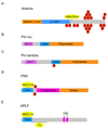
Domain boundaries and major features of Artemis, PNK, APLF, pol μ and pol λ are represented as in Figure 2. (A) Artemis: The metallo β lactamase domain (aa 1–155) and β-CASP domain (aa 156–385) are as described in [86]. Amino acids 398–403 are required for interaction with DNA-PKcs [89, 95]. The C-terminal region of Artemis is highly phosphorylated at multiple site both in vitro and in vivo [62, 65, 89, 90, 92, 93] but the effects of phosphorylation on function are not known. (B and C) Pol μ and λ: Domain boundaries for the lyase (aa. 156–227) and polymerase (aa 227–494) domains of pol mu are based on a structure based alignment of pol X family members [103, 182]. The BRCT (aa. 29–109) domain is as described in the NCBI database. The lyase (aa 242–327), polymerase (aa 327–575) and the BRCT domains (aa 35–125) of pol lambda are from [103, 182]. (D) PNK: Domain boundaries for the FHA (aa 6–110), phosphatase (aa 145–337) and kinase (aa 341–521) domains are based on the X-ray crystal structure [183]. CK2 phosphorylated XRCC4 and XRCC1 interact with the FHA domain [109, 174]. Serines 114 and 126 are IR-induced phosphorylated sites of unknown function [70]. (E) APLF: Amino acids 20–102 compose the FHA domain. XRCC1/XRCC4 bind within the FHA domain [113, 114]. The poly(ADP-ribose) binding zinc finger (PBZ) regions (aa 377–398 and aa 419–440) of APLF are shown in purple [115]. Although APLF has endonuclease and exonuclease activity, these domains have yet to be defined.
Figure 5
Figure 5. Major features of DNA ligase IV, XRCC4 and XLF
Domain boundaries and major features of DNA ligase IV, XRCC4 and XLF are represented as in Figure 2. (A) XRCC4: The head (aa 1–115) and stalk domains (aa 135–233) of XRCC4 are based on the x-ray crystal structure [184]. The region of XRCC4 required for dimerization (aa 119–155) is from [132]; DNA ligase IV interacts with XRCC4 between amino acids 173–195 [134]; CK-2 phosphorylates XRCC4 at threonine 233 [109] and DNA-PK phosphorylates XRCC4 at serine 260 and serine 318 in vitro [58]. The XLF binding site (aa 63–99) is from [150]. (B) DNA ligase IV: The N-terminal (aa. 14–203) and ligase domain (aa. 248–451) boundaries are as found in the NCBI database. The XRCC4 binding site in DNA ligase IV (aa. 755–782; [134]) is located between the BRCT domains (aa. 661–731 and aa. 829–898); In vitro, DNA ligase IV is phosphorylated at threonine 650, serine 668 and serine 672 [64]. (C) XLF: The head (aa 1–135) and stalk (aa 135–233) domains are based on the X-ray crystal structure [184]. Leucine 115 is crucial for XRCC4 binding and amino acids 125–224 are involved in homodimerization [150, 184]. In vivo, XLF is phosphorylated at serine 245 by DNA-PK and serine 251 by ATM but the effect of phosphorylation on function is unknown [59].

References

    1. Povirk LF. Biochemical mechanisms of chromosomal translocations resulting from DNA double-strand breaks. DNA Repair (Amst) 2006;5:1199–1212. - PubMed
    1. Helleday T, Lo J, van Gent DC, Engelward BP. DNA double-strand break repair: from mechanistic understanding to cancer treatment. DNA Repair (Amst) 2007;6:923–935. - PubMed
    1. Kastan MB, Bartek J. Cell-cycle checkpoints and cancer. Nature. 2004;432:316–323. - PubMed
    1. Lobrich M, Jeggo PA. The impact of a negligent G2/M checkpoint on genomic instability and cancer induction. Nat Rev Cancer. 2007;7:861–869. - PubMed
    1. Hoeijmakers JH. Genome maintenance mechanisms for preventing cancer. Nature. 2001;411:366–374. - PubMed

Publication types

Substances