. 2009 Jan 9;28(1):5.
doi: 10.1186/1756-9966-28-5.

Tissue microarray analysis of eIF4E and its downstream effector proteins in human breast cancer

Affiliations

Tissue microarray analysis of eIF4E and its downstream effector proteins in human breast cancer

Heather E Kleiner et al. J Exp Clin Cancer Res. .

Erratum in

  • J Exp Clin Cancer Res. 2009;28:54

Abstract

Background: Eukaryotic initiation factor 4E (eIF4E) is elevated in many cancers and is a prognostic indicator in breast cancer. Many pro-tumorigenic proteins are selectively translated via eIF4E, including c-Myc, cyclin D1, ornithine decarboxylase (ODC), vascular endothelial growth factor (VEGF) and Tousled-like kinase 1B (TLK1B). However, western blot analysis of these factors in human breast cancer has been limited by the availability of fresh frozen tissue and the labor-intensive nature of the multiple assays required. Our goal was to validate whether formalin-fixed, paraffin-embedded tissues arranged in a tissue microarray (TMA) format would be more efficient than the use of fresh-frozen tissue and western blot to test multiple downstream gene products.

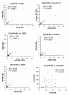
Results: Breast tumor TMAs were stained immunohistochemically and quantitated using the ARIOL imaging system. In the TMAs, eIF4E levels correlated strongly with c-Myc, cyclin D1, TLK1B, VEGF, and ODC. Western blot comparisons of eIF4E vs. TLK1B were consistent with the immunohistochemical results. Consistent with our previous western blot results, eIF4E did not correlate with node status, ER, PR, or HER-2/neu.

Conclusion: We conclude that the TMA technique yields similar results as the western blot technique and can be more efficient and thorough in the evaluation of several products downstream of eIF4E.


Figures

Figure 1
Figure 1
Low magnification (100 ×) of human breast cancer specimens in TMA3 stained immunohistochemically for ODC. Boxes indicate specimen type. The specimens marked "low 4E" and "high 4E" are also shown in Figure 3. The top right corner of the TMA was left blank, and a pancreas specimen was placed in the lower right hand corner of the TMA in order to verify proper orientation. IDC, intraductal carcinoma; DCIS, ductal carcinoma in situ.
Figure 2
Figure 2
High magnification (400 ×) of human breast cancer specimen from TMA3 stained immunohistochemically for ODC. Note the predominantly cytosolic staining of ODC, whereas the nuclei were counterstained blue.
Figure 3
Figure 3
Representative example of human breast cancer specimens from TMA3 that expressed either low (left panel) or high (right panel) eIF4E. Matching specimens from the same patient are shown for c-Myc, cyclin D1, ODC, TLK1B, and VEGF (200 × magnification).
Figure 4
Figure 4
Correlation of immunohistochemical expression of eIF4E vs c-Myc [A], cyclin D1 [B], ODC [C], TLK1B [D], VEGF [E] from TMA3. Figures represent the integrated optical density (IOD) of immunohistochemical staining intensity normalized to cytokeratin. Protein expression of eIF4E and TLK1B were also compared by western blot analysis [F], in which values represent expression of eIF4E and TLK1B as fold- over benign. All comparisons were done using Spearman's rank correlation. Rho- and p- values for each comparison are displayed in each panel.

References

    1. Jemal A, Siegel R, Ward E, Murray T, Xu J, Thun MJ. Cancer statistics, 2007. CA Cancer J Clin. 2007;57:43–66. - PubMed
    1. De Benedetti A, Harris AL. eIF4E expression in tumors: its possible role in progression of malignancies. Int J Biochem Cell Biol. 1999;31:59–72. doi: 10.1016/S1357-2725(98)00132-0. - DOI - PubMed
    1. Dillon RL, White DE, Muller WJ. The phosphatidyl inositol 3-kinase signaling network: implications for human breast cancer. Oncogene. 2007;26:1338–1345. doi: 10.1038/sj.onc.1210202. - DOI - PubMed
    1. Santen RJ, Song RX, McPherson R, Kumar R, Adam L, Jeng MH, Yue W. The role of mitogen-activated protein (MAP) kinase in breast cancer. J Steroid Biochem Mol Biol. 2002;80:239–256. doi: 10.1016/S0960-0760(01)00189-3. - DOI - PubMed
    1. Wu JT, Kral JG. The NF-kappaB/IkappaB signaling system: a molecular target in breast cancer therapy. J Surg Res. 2005;123:158–169. doi: 10.1016/j.jss.2004.06.006. - DOI - PubMed

Publication types

MeSH terms

Substances