. 2009 Jan;96(1):35-44.
doi: 10.1016/j.bpj.2008.09.027.

A CaV1.1 Ca2+ channel splice variant with high conductance and voltage-sensitivity alters EC coupling in developing skeletal muscle

Affiliations

A CaV1.1 Ca2+ channel splice variant with high conductance and voltage-sensitivity alters EC coupling in developing skeletal muscle

Petronel Tuluc et al. Biophys J. 2009 Jan.

Abstract

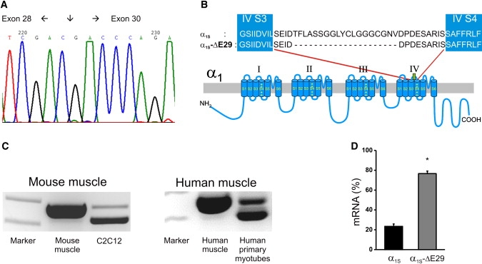
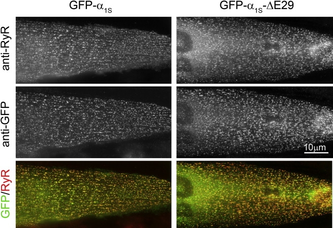
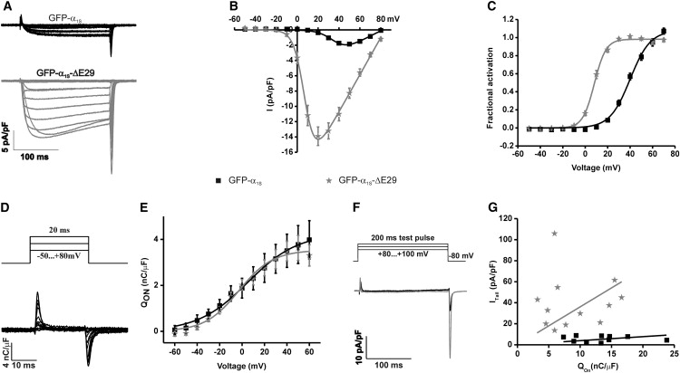
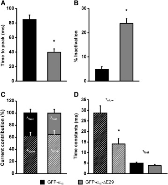
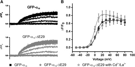
The Ca(2+) channel alpha(1S) subunit (Ca(V)1.1) is the voltage sensor in skeletal muscle excitation-contraction (EC) coupling. Upon membrane depolarization, this sensor rapidly triggers Ca(2+) release from internal stores and conducts a slowly activating Ca(2+) current. However, this Ca(2+) current is not essential for skeletal muscle EC coupling. Here, we identified a Ca(V)1.1 splice variant with greatly distinct current properties. The variant of the CACNA1S gene lacking exon 29 was expressed at low levels in differentiated human and mouse muscle, and up to 80% in myotubes. To test its biophysical properties, we deleted exon 29 in a green fluorescent protein (GFP)-tagged alpha(1S) subunit and expressed it in dysgenic (alpha(1S)-null) myotubes. GFP-alpha(1S)Delta 29 was correctly targeted into triads and supported skeletal muscle EC coupling. However, the Ca(2+) currents through GFP-alpha(1S)Delta 29 showed a 30-mV left-shifted voltage dependence of activation and a substantially increased open probability, giving rise to an eightfold increased current density. This robust Ca(2+) influx contributed substantially to the depolarization-induced Ca(2+) transient that triggers contraction. Moreover, deletion of exon 29 accelerated current kinetics independent of the auxiliary alpha(2)delta-1 subunit. Thus, characterizing the Ca(V)1.1 Delta 29 splice variant revealed the structural bases underlying the specific gating properties of skeletal muscle Ca(2+) channels, and it suggests the existence of a distinct mode of EC coupling in developing muscle.


Figures

Figure 1
Figure 1
Detection of GFP-α1SΔ29 in human and mouse myotubes. (A) Sequence of the boundary between exons 28 and 30. (B) Location of exon 29 in a domain model of CaV1.1. (C) Full-length (upper band) and CaV1.1Δ29 (lower band) detected by RT-PCR amplification of exons 26–30 in RNA prepared from mouse muscle, C2C12 myotubes, human muscle, and human primary myotubes. (D) The fraction of CaV1.1 transcripts with and without exon 29 measured with quantitative RT-PCR in mRNA from mouse myotubes. Error bars represent the mean ± SE, p << 0.001.
Figure 2
Figure 2
GFP-α1SΔ29 is targeted into junctions between the SR and t-tubules or the plasma membrane. Dysgenic myotubes expressing full-length GFP-α1S (left) or the GFP-α1SΔ29 splice variant (right) were double-immunolabeled with anti-RyR (upper) and anti-GFP (middle). Clusters of GFP-α1S and GFP-α1SΔ29 colocalized with the RyR1 (lower, yellow clusters in color overlay) indicate the correct targeting of both Ca2+ channel variants into t-tubule/SR or plasma membrane/SR junctions. Scale bar, 10 μm.
Figure 3
Figure 3
GFP-α1SΔ29 has increased current density, voltage sensitivity of activation, and open probability (Po). (A) Representative whole-cell currents from myotubes expressing either GFP-α1S or GFP-α1SΔ29. (B) I/V curves show that the peak current density of GFP-α1SΔ29 is increased. (C) Voltage dependence of activation is shifted toward more negative potentials for GFP-α1SΔ29 compared to full-length GFP-α1S. (D and E) Analysis of the “On” gating charges (Qon) while currents are blocked with Cd2+/ La3+ shows that deletion of exon 29 did not alter the expression of functional channels in the membrane. (F and G) The amplitudes of the tail currents, recorded at the reversal potential were plotted against Qon. The increased slope of the linear regression indicates that the channel Po is considerably increased in GFP-α1SΔ29 compared to GFP-α1S. Error bars indicate the SE.
Figure 4
Figure 4
Deletion of exon 29 accelerates Ca2+ current kinetics. (A and B) Currents recorded from myotubes expressing GFP-α1SΔ29 exhibit a significantly shorter time to peak (A) and an increased fractional inactivation during a 200-ms pulse (B). The rising phase of Ca2+ currents was fitted by a double-exponential function and the amplitudes and time constants of the two components were calculated. Neither the ratio between fast and slow components (C) nor the time constant of the fast component (D, solid bars) was affected by the deletion of exon 29. The time constant of the slow component was significantly faster in GFP-α1SΔ29 compared to GFP-α1S (D, hatched bars). Error bars represent the mean ± SE.
Figure 5
Figure 5
Deletion of exon 29 does not affect the interaction of α1 with the α2δ-1 subunit. The α2δ-1 subunit was depleted with shRNA in myotubes expressing GFP-α1SΔ29. This resulted in a further acceleration of activation and inactivation kinetics as seen in the sample recordings (A). The time to peak was further reduced (B), and the percentage of inactivation was increased (C). Kinetic analysis of the activation phase revealed only one component of activation (D, white bar) with a time constant equal to τfast in GFP-α1SΔ29 controls (E, gray bars), indicating the loss of the slow-activating component. Error bars represent the mean ± SE.
Figure 6
Figure 6
GFP-α1SΔ29 supports skeletal muscle type EC coupling with an additional component of Ca2+ influx. Depolarization-induced Ca2+ transients were recorded in dysgenic myotubes reconstituted with GFP-α1S (A, upper) or GFP-α1SΔ29 (A, lower). The voltage dependence of activation was not altered by the deletion of exon 29, but Ca2+ transients were augmented by a component that declined at voltages near the reversal potential and could be inhibited by blocking Ca2+ currents with Cd2+/La3+ (B). Error bars represent the mean ± SE.

References

    1. Melzer W., Herrmann-Frank A., Luttgau H.C. The role of Ca2+ ions in excitation-contraction coupling of skeletal muscle fibres. Biochim. Biophys. Acta. 1995;1241:59–116. - PubMed
    1. Jurkat-Rott K., Lehmann-Horn F. The impact of splice isoforms on voltage-gated calcium channel α1 subunits. J. Physiol. 2004;554:609–619. - PMC - PubMed
    1. Abernethy D.R., Soldatov N.M. Structure-functional diversity of human L-type Ca2+ channel: perspectives for new pharmacological targets. J. Pharmacol. Exp. Ther. 2002;300:724–728. - PubMed
    1. Diebold R.J., Koch W.J., Ellinor P.T., Wang J.J., Muthuchamy M. Mutually exclusive exon splicing of the cardiac calcium channel α1 subunit gene generates developmentally regulated isoforms in the rat heart. Proc. Natl. Acad. Sci. USA. 1992;89:1497–1501. - PMC - PubMed
    1. Perez-Reyes E., Wei X.Y., Castellano A., Birnbaumer L. Molecular diversity of L-type calcium channels. Evidence for alternative splicing of the transcripts of three non-allelic genes. J. Biol. Chem. 1990;265:20430–20436. - PubMed

Publication types

MeSH terms