

Fate plasticity of adult hippocampal progenitors: biological relevance and therapeutic use
- PMID: 19135265
- PMCID: PMC2752320
- DOI: 10.1016/j.tips.2008.11.003
Fate plasticity of adult hippocampal progenitors: biological relevance and therapeutic use
Abstract
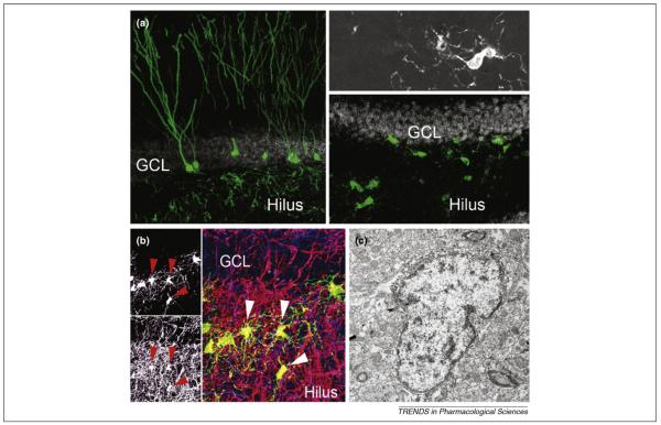
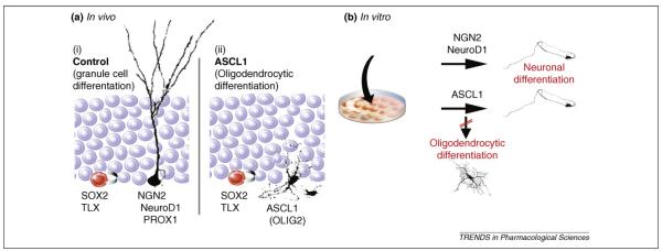
Adult hippocampal stem/progenitor cells (AHPs) continuously give rise to new neurons throughout life, which might be an important determinant of hippocampus-dependent function. Strikingly, the fate potential of AHPs is not restricted to the neuronal lineage because AHPs can be genetically induced to generate oligodendrocytes within their in vivo niche by AHP-specific ectopic expression of the basic-helix-loop-helix (bHLH) transcription factor achaete-scute complex-like 1 (ASCL1). Fate plasticity of AHPs is controlled by cell-autonomous and also niche-dependent mechanisms. Here, we discuss the biological importance and potential therapeutic applications of retained fate plasticity of AHPs in the adult mammalian brain in addition to the future scientific inquiries indicated by this finding.
Figures
Similar articles
-
Directed differentiation of hippocampal stem/progenitor cells in the adult brain.Nat Neurosci. 2008 Aug;11(8):888-93. doi: 10.1038/nn.2148. Epub 2008 Jun 29. Nat Neurosci. 2008. PMID: 18587391 Free PMC article.
-
Ascl1 defines sequentially generated lineage-restricted neuronal and oligodendrocyte precursor cells in the spinal cord.Development. 2007 Jan;134(2):285-93. doi: 10.1242/dev.02727. Epub 2006 Dec 13. Development. 2007. PMID: 17166924
-
Ascl1 (Mash1) lineage cells contribute to discrete cell populations in CNS architecture.Mol Cell Neurosci. 2008 Aug;38(4):595-606. doi: 10.1016/j.mcn.2008.05.008. Epub 2008 May 20. Mol Cell Neurosci. 2008. PMID: 18585058 Free PMC article.
-
Neurogenesis and hippocampal plasticity in adult brain.Curr Top Behav Neurosci. 2013;15:31-48. doi: 10.1007/7854_2012_217. Curr Top Behav Neurosci. 2013. PMID: 22879073 Review.
-
Synapse formation on adult-born hippocampal neurons.Eur J Neurosci. 2011 Mar;33(6):1062-8. doi: 10.1111/j.1460-9568.2011.07604.x. Eur J Neurosci. 2011. PMID: 21395849 Review.
Cited by
-
The roles of BDNF, pCREB and Wnt3a in the latent period preceding activation of progenitor cell mitosis in the adult dentate gyrus by fluoxetine.PLoS One. 2010 Oct 27;5(10):e13652. doi: 10.1371/journal.pone.0013652. PLoS One. 2010. PMID: 21048974 Free PMC article.
-
Contribution of constitutively proliferating precursor cell subtypes to dentate neurogenesis after cortical infarcts.BMC Neurosci. 2010 Nov 17;11:146. doi: 10.1186/1471-2202-11-146. BMC Neurosci. 2010. PMID: 21083887 Free PMC article.
-
Engineering of Adult Neurogenesis and Gliogenesis.Cold Spring Harb Perspect Biol. 2016 May 2;8(5):a018861. doi: 10.1101/cshperspect.a018861. Cold Spring Harb Perspect Biol. 2016. PMID: 27091941 Free PMC article. Review.
-
Allopregnanolone reverses neurogenic and cognitive deficits in mouse model of Alzheimer's disease.Proc Natl Acad Sci U S A. 2010 Apr 6;107(14):6498-503. doi: 10.1073/pnas.1001422107. Epub 2010 Mar 15. Proc Natl Acad Sci U S A. 2010. PMID: 20231471 Free PMC article.
-
Neuroprotective effects of dexmedetomidine against hyperoxia-induced injury in the developing rat brain.PLoS One. 2017 Feb 3;12(2):e0171498. doi: 10.1371/journal.pone.0171498. eCollection 2017. PLoS One. 2017. PMID: 28158247 Free PMC article.
References
-
- Gage FH. Mammalian neural stem cells. Science. 2000;287:1433–1438. - PubMed
-
- Zhao C, et al. Mechanisms and functional implications of adult neurogenesis. Cell. 2008;132:645–660. - PubMed
-
- Goldman SA. Disease targets and strategies for the therapeutic modulation of endogenous neural stem and progenitor cells. Clin. Pharmacol. Ther. 2007;82:453–460. - PubMed
-
- Kempermann G, et al. Milestones of neuronal development in the adult hippocampus. Trends Neurosci. 2004;27:447–452. - PubMed
Publication types
MeSH terms
Substances
Grants and funding
LinkOut - more resources
Full Text Sources
Other Literature Sources
