. 2009 Feb 20;386(2):504-19.
doi: 10.1016/j.jmb.2008.12.043. Epub 2008 Dec 25.

The Escherichia coli cell division protein and model Tat substrate SufI (FtsP) localizes to the septal ring and has a multicopper oxidase-like structure

Affiliations

The Escherichia coli cell division protein and model Tat substrate SufI (FtsP) localizes to the septal ring and has a multicopper oxidase-like structure

Michael Tarry et al. J Mol Biol. .

Abstract

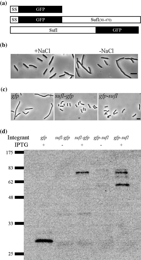
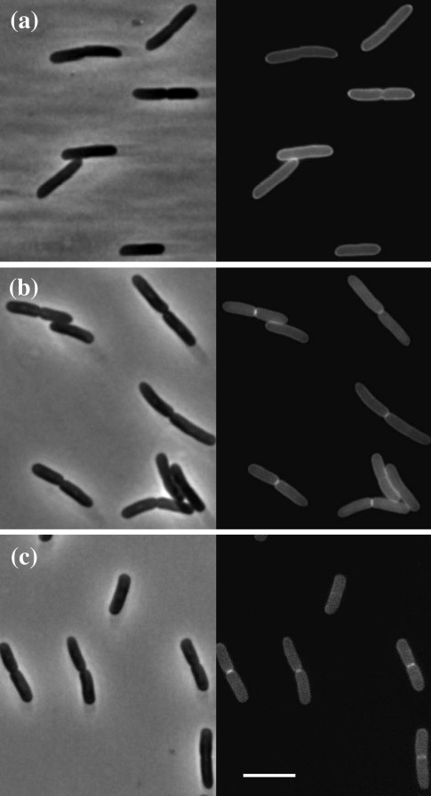
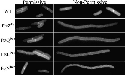
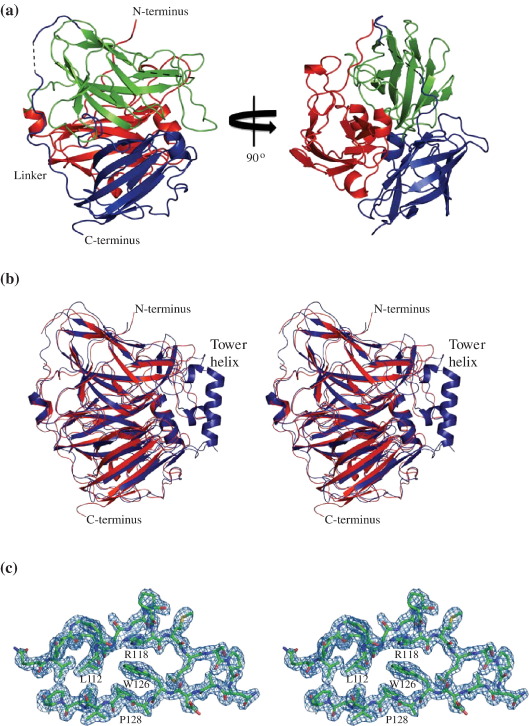
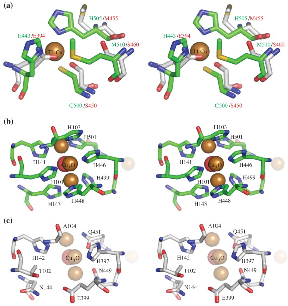
The Escherichia coli protein SufI (FtsP) has recently been proposed to be a component of the cell division apparatus. The SufI protein is also in widespread experimental use as a model substrate in studies of the Tat (twin arginine translocation) protein transport system. We have used SufI-GFP (green fluorescent protein) fusions to show that SufI localizes to the septal ring in the dividing cell. We have also determined the structure of SufI by X-ray crystallography to a resolution of 1.9 A. SufI is structurally related to the multicopper oxidase superfamily but lacks metal cofactors. The structure of SufI suggests it serves a scaffolding rather than an enzymatic role in the septal ring and reveals regions of the protein likely to be involved in the protein-protein interactions required to assemble SufI at the septal ring.


Figures

Fig. 1
Fig. 1
(a) Cartoon of gfp constructs. “SS” refers to a Tat signal sequence from TorA. SufI is a Tat substrate and therefore does not need an artificial signal sequence. (b) A sufI null mutant has mild division defects. EC1751 (sufI<>aph) was grown at 37 °C in LB with or without 1% NaCl, fixed, and photographed under phase contrast microscopy. The scale bar represents 5 μm. (c) GFP/SufI fusions rescue division defects. Derivatives of EC1751 (sufI<>aph) expressing the indicated gfp construct were grown in LB0N and photographed as above. The strains shown are EC1874, EC1875 and EC1876. The scale bar represents 5 μm. (d) Immunoblot of strains that express gfp, sufI-gfp or gfp-sufI under the control of an IPTG-inducible promoter. Sample concentrations were normalized by OD600, except that fourfold more of the sufI-gfp sample was loaded because this fusion gene is not highly expressed. The blot was probed with anti-GFP serum. The strains shown are gfp (EC1874), sufI-gfp (EC1876) and gfp-sufI (EC1875). Molecular mass markers are indicated at the left.
Fig. 2
Fig. 2
Septal localization of SufI. Phase (left) and fluorescence (right) micrographs of an sufI<>aph null mutant expressing (a) gfp, (b) gfp-sufI and (c) sufI-gfp. The scale bar represents 5 μm. The strains shown are EC1874, EC1875 and EC1876. They were grown in LB with 1% NaCl and 1 mM IPTG at 30 °C.
Fig. 3
Fig. 3
Recruitment of SufI to septal rings requires other Fts proteins. The top two panel sets show a wild-type (WT) strain or ftsZ(Ts) mutant grown at 30 °C in LB (permissive) or 37 °C in LB0N (non-permissive). The strains shown are EC1873 and EC2065, both of which carry a chromosomal sufI-gfp fusion. Traditionally, 42 °C has been used as the non-permissive temperature for ftsZ84(Ts), but we found that SufI-GFP was not fluorescent at 42 °C. The bottom three panel sets show FtsQ, FtsL or FtsN depletion strains. The strains shown are JM265, JOE170 and EC1908 and carry the sufI-gfp plasmid pDSW932. These strains produce the indicated Fts protein from an arabinose-dependent promoter and were grown in the presence of arabinose (permissive) or glucose (non-permissive). The cells shown are representative. For quantitative data, see Supplemental Table 1.
Fig. 4
Fig. 4
Structure of SufI. (a) Cartoon representation of SufI. The structure shown is for chain A of the orthorhombic space group with domain 1 shown in red, domain 2 in green and domain 3 in blue. Regions of missing density are shown as black dotted lines. The arrow in the region of missing density indicates where the protein is subject to proteolytic cleavage. (b) Stereo view showing CueO (blue) overlaid on the orthorhombic SufI (red) structure. PDB ID 1KV7 was overlaid onto the orthorhombic chain A of SufI with the program CCP4-Lsqkab. The positions of the N- and C-termini of the proteins are shown and the tower region of CueO is labelled for reference. The orientation is as in (a). (c) Stereo view showing representative electron density (2Fo − Fc) of orthorhombic chain A, residues 107–131 contoured at 1σ. The positions of the invariant residues leucine 112, arginine 118, tryptophan 126 and proline 128 are labelled. The images were generated with Pymol.
Fig. 5
Fig. 5
Comparison of the copper-binding sites of CueO and the equivalent regions of SufI. (a) Stereo view of the copper-binding residues of the T1 copper-binding site of CueO (green sticks) overlaid on the corresponding region of SufI (white sticks). The structure of CueO (PDB ID 1KV7) was overlaid on the orthorhombic chain A of SufI with the program CCP4-Lsqkab. The residues are labelled in green for CueO and red for SufI. The T1 copper of CueO is shown as a space-filling sphere. (b) Stereo view representation of the trinuclear copper-binding site of CueO showing the copper-binding residues as stick models. The trinuclear centre is shown as space-filling spheres. (c) The corresponding residues of SufI shown as sticks with the corresponding position of the trinuclear centre of CueO shown as transparent space-filling spheres. The structure of CueO (PDB ID 1KV7) was overlaid on the orthorhombic chain A of SufI with the program CCP4-Lsqkab. The images were generated with Pymol.
Fig. 6
Fig. 6
Sequence alignment of SufI proteins with CueO. Putative SufI sequences from various bacteria were identified with the computer program BLAST. The SufI sequences were aligned with one another and also with CueO from E. coli with ClustalW and coloured with WebESPript. Key: white character inside red box, strict identity; red character, similarity across SufI sequences; blue frame, similarity between conserved SufI sequences and CueO; orange box, differences between conserved SufI sequences and CueO. Two regions identified as having potential roles in SufI function are highlighted in green. Secondary structural elements for E. coli SufI and CueO are indicated above and below their respective sequences. Yellow triangles identify the type 1 copper centre coordinating residues in CueO, while blue triangles identify the residues in CueO that coordinate to the trinuclear copper centre. Eco, Escherichia coli; Shi, Shigella flexneri; Sal, Salmonella typhimurium; Ent, Enterobacter sp. 638; Yer, Yersinia enterocolitica; Erw, Erwinia carotovora; Sod, Sodalis glossinidius; Pho, Photorhabdus luminescens; Man, Mannheimia succiniciproducens; Hae, Haemophilus influenzae; Pas, Pasteurella multocida; Act, Actinobacillus pleuropneumonia.
Fig. 7
Fig. 7
Identification and localization of conserved residues in SufI. (a) CueO: the conserved residues Gly113 and Gly114 (green sticks) sit above the trinuclear copper centre (shown as transparent spheres). Residues Gly117, Arg125 and Val127 (blue sticks) form the brim of a surface cavity that exposes the 112–114 loop (DGG in CueO, conserved as DGX across all multicopper oxidases) to the solvent. (b) SufI: in green, the glycine residues Gly114 and Gly115 corresponding to the glycines in (a); in blue, the residues Arg118, Trp126 and Pro128, which are all highly conserved across SufI sequences and prevent solvent access to the 112–114 loop in the SufI structure. (c) A surface representation of SufI, coloured red (surface residue most conserved) through orange, yellow, and green, to blue (least conserved). The N- and C-termini of the protein are shown for reference and highly conserved surface residues are labelled. A black star marks the location of the residues 118–128 that cover the pocket corresponding to the CueO catalytic centre. The right-hand panel shows a second view of the SufI surface, rotated with respect to the left-hand panel by 180° around the vertical axis. Images were generated using Pymol with (c) drawn using a PDB file produced by the WHISCY server.

References

    1. Arends S.J.R., Williams K.B., Kustusch R.J., Weiss D.S., Erhmann M. The Periplasm. ASM Press; Washington, DC: 2007. Cell division; pp. 173–197.
    1. Errington J., Daniel R.A., Scheffers D.J. Cytokinesis in bacteria. Microbiol. Mol. Biol. Rev. 2003;67:52–65. table. - PMC - PubMed
    1. Goehring N.W., Beckwith J. Diverse paths to midcell: assembly of the bacterial cell division machinery. Curr. Biol. 2005;15:R514–R526. - PubMed
    1. Margolin W. FtsZ and the division of prokaryotic cells and organelles. Nat. Rev. Mol. Cell. Biol. 2005;6:862–871. - PMC - PubMed
    1. Vicente M., Rico A.I., Martinez-Arteaga R., Mingorance J. Septum enlightenment: assembly of bacterial division proteins. J. Bacteriol. 2006;188:19–27. - PMC - PubMed

Publication types

MeSH terms

Substances

LinkOut - more resources