. 2009 Mar;142(1-2):59-67.
doi: 10.1016/j.pain.2008.11.013. Epub 2009 Jan 9.

Periganglionic inflammation elicits a distally radiating pain hypersensitivity by promoting COX-2 induction in the dorsal root ganglion

Affiliations

Periganglionic inflammation elicits a distally radiating pain hypersensitivity by promoting COX-2 induction in the dorsal root ganglion

Fumimasa Amaya et al. Pain. 2009 Mar.

Abstract

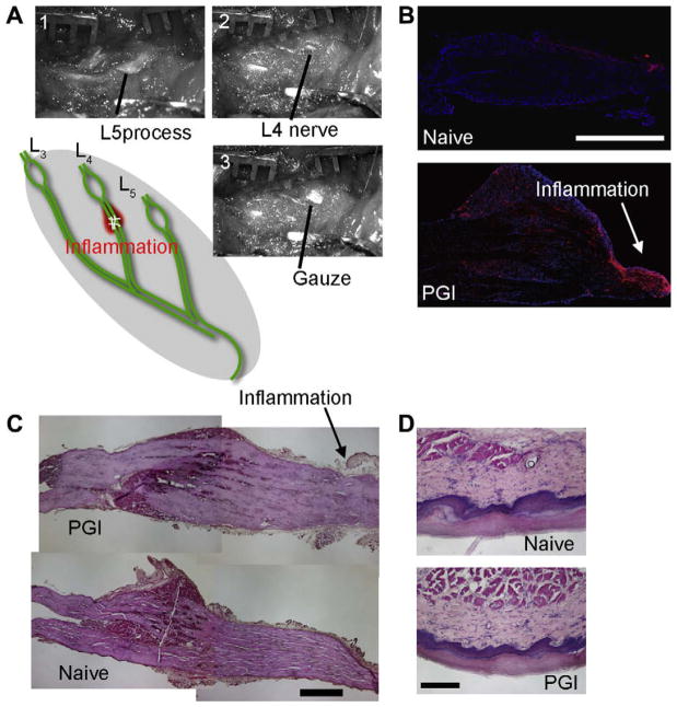
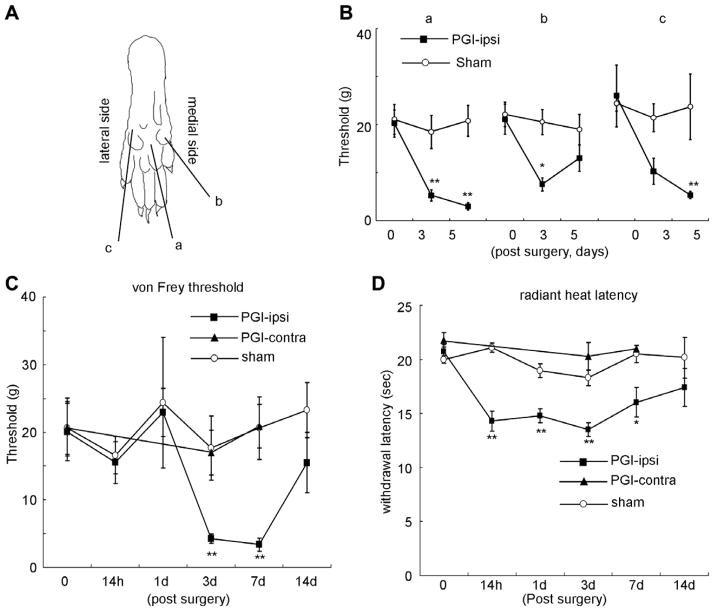
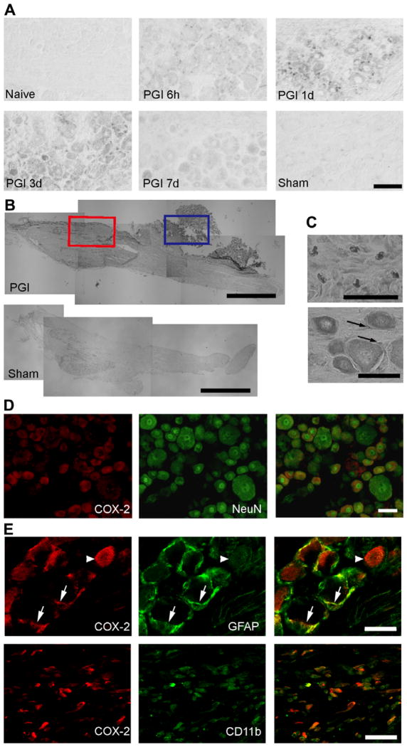
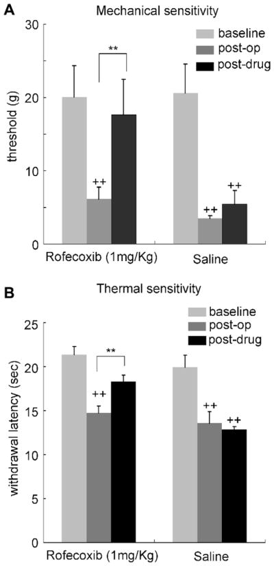
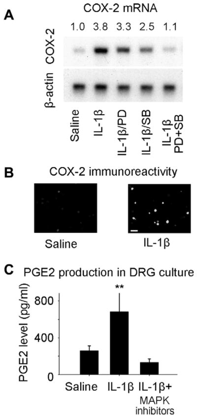
We have developed a model in which inflammation contiguous to and within a dorsal root ganglion (DRG) was generated by local application of complete Freund's adjuvant (CFA) to the L4 lumbar spinal nerve as it exits from the intervertebral foramen. The periganglionic inflammation (PGI) elicited a marked reduction in withdrawal threshold to mechanical stimuli and an increase in heat pain sensitivity in the ipsilateral hindpaw in the absence of any hindpaw inflammation. The pain sensitivity appeared within hours and lasted for a week. The PGI also induced a prominent increase in IL-1beta and TNF-alpha levels in the DRG and of cyclooxygenase-2 (COX-2) expression in neurons and satellite cells. A selective COX-2 inhibitor reduced the PGI-induced hyperalgesia. We also show that IL-1beta induces COX-2 expression and prostaglandin release in DRG neurons in vitro in a MAP kinase-dependent fashion. The COX-2 induction was prevented by ERK and p38 inhibitors. We conclude that periganglionic inflammation increases cytokine levels, including IL-1beta, leading to the transcription of COX-2 and prostaglandin production in the affected DRG, and thereby to the development of a dermatomally distributed pain hypersensitivity.


Conflict of interest statement

Conflict of interest

The authors do not have a conflict of interest. T.S. is currently employed by Wyeth.

Figures

Fig. 1
Fig. 1
Periganglionic inflammation (PGI). (A) Procedure for surgical treatment of PGI. After identifying L5 process (A-1), the process was cut to visualize L4 nerve (A-2). Cotton gauze with CFA or saline was put onto the nerve (A-3). Acute inflammation of the L4 spinal nerve produced by CFA application immediately distal to the L4 DRG. (B) Immunohistochemistry for CD11b, a marker for activated macrophages shows the accumulation of immune cells at the site of the CFA application and in the DRG. Red CD11b, blue DAPI nuclear staining. CD11b positive cells were not observed in DRGs from naive animals. (C) Hematoxylin–Eosin (HE) staining of the DRG from PGI animals. No sign of mechanical compression was observed. Arrows indicate inflammation site. Scale bar = 500 μm. (D) HE staining of the skin of the hindpaw from PGI and naive animals. No inflammatory reaction (inflammatory cell accumulation or tissue edema) was observed in the paw. Scale bar = 200 μm.
Fig. 2
Fig. 2
Pain hypersensitivity in the hindpaw following PGI. (A) Sites tested on the paw surface are indicated by a, b, and c. (B) Mechanical thresholds (von Frey) of these three sites on the ipsilateral plantar surface after PGI. (C) Time course of the change in mechanical threshold in area a. (D) Changes in withdrawal time to a noxious radiant heat stimulus of the ipsilateral hindpaw after PGI. N = 7 for each group. *p < 0.05 vs. sham operation group. **p < 0.01 vs. sham operation group.
Fig. 3
Fig. 3
COX-2 and inflammatory cytokine induction in the DRG following PGI. (A) COX-2 mRNA levels measured in L3–L5 DRGs, 3 days after PGI and compared to a sham operation or hindpaw inflammation. (B) Hindpaw inflammation but not PGI-induced COX-2 mRNA in the dorsal horn (DH). (C) Increase in IL-1β mRNA in L4 DRG of PGI treated animals. (D) Increase in TNF-α mRNA in L4 DRG of PGI treated animals. *p < 0.05 vs. sham control. **p < 0.01 vs. sham control. N = 4 for each group of animals.
Fig. 4
Fig. 4
COX-2 expression in the DRG after PGI. (A) COX-2 mRNA detected by in situ hybridization. COX-2 mRNA is first detected in small cells 6 h after the periganglionic CFA treatment. Three days after treatment, relatively strong COX-2 expression is observed in neurons. Scale bar = 50 μm. (B) Immunohistochemical study showing the distribution of COX-2 positive cells in the DRG 3 days after the PGI treatment, compared to sham. COX-2 positive cells are detected within (red square) and adjacent to the DRG (blue square). Scale bar = 1000 μm. (C) Higher magnification of two areas in B. Top: COX-2 expression observed in the connective tissue (area in the blue square in B). Bottom: COX-2 expression in neuronal and satellite cells in the DRG (area in the red square in B). Arrows indicate COX-2 positive satellite cells. Scale bar = 50 μm. (D) Neuronal COX-2 expression. Double immunohistochemistry for COX-2 (red) with NeuN, neuronal cell marker (green) revealed that COX-2 co-localizes with the neuronal cell marker NeuN in the DRG. Scale bar = 1200 μm. (E) Non-neuronal COX-2 expression. A single confocal optical section analysis of COX-2 expression (red) with GFAP (green) or CD11b (green) positive cells. COX-2 expression co-localizes with GFAP and CD11b. The COX-2/GFAP images were taken within the DRG, while the COX-2/CD11b images were taken in an area adjacent to the DRG. Scale bar = 50 μm.
Fig. 5
Fig. 5
A selective COX-2 inhibitor rofecoxib (1 mg/kg administrated subcutaneously) reduced the mechanical (A) and thermal (B) pain hypersensitivity produced by PGI. Behavioral analysis was performed 3 days after the PGI procedure, and the effect of rofecoxib was assessed 30 min after administration. N = 7 in each group. ++: p < 0.01 vs. pre-treatment baseline. **p < 0.01 vs. saline injected control.
Fig. 6
Fig. 6
(A) Signal pathways involved in COX-2 induction in DRG neurons cultured from na rats 6 h after exposure to IL-1β. (A) Application of IL-1β to cultured DRG neurons produced an increase in COX-2 mRNA that was partially blocked by administration either of SB203580, a p38 MAP kinase inhibitor or of PD98059, an ERK kinase inhibitor. Co-application of the p38/ERK inhibitors blocked COX-2 induction almost completely. The COX-2/β-actin signal density ratio is presented above the image. (B) COX-2 immunoreactivity in cultured DRG neurons after IL-1β. Scale bar = 50 μm (C) Prostaglandin levels in the media of cultured DRG neurons were increased by IL-1β in a p38/ERK dependent fashion.

Comment in

  • What causes low back pain?
    Devor M, Tal M. Devor M, et al. Pain. 2009 Mar;142(1-2):11-2. doi: 10.1016/j.pain.2009.01.002. Epub 2009 Jan 31. Pain. 2009. PMID: 19181449 No abstract available.

References

    1. Amaya F, Oh-hashi K, Naruse Y, Iijima N, Ueda M, Shimosato G, et al. Local inflammation increases vanilloid receptor 1 expression within distinct subgroups of DRG neurons. Brain Res. 2003;963:190–6. - PubMed
    1. Amaya F, Wang H, Costigan M, Allchorne AJ, Hatcher JP, Egerton J, et al. The voltage-gated sodium channel Na(v)1 9 is an effector of peripheral inflammatory pain hypersensitivity. J Neurosci. 2006;26:12852–60. - PMC - PubMed
    1. Andersson GB. Epidemiological features of chronic low-back pain. Lancet. 1999;354:581–5. - PubMed
    1. Appleton I, Tomlinson A, Mitchell JA, Willoughby DA. Distribution of cyclooxygenase isoforms in murine chronic granulomatous inflammation. Implications for future anti-inflammatory therapy. J Pathol. 1995;176:413–20. - PubMed
    1. Arnau JM, Vallano A, Lopez A, Pellise F, Delgado MJ, Prat N. A critical review of guidelines for low back pain treatment. Eur Spine J. 2006;15:543–53. - PMC - PubMed

Publication types

MeSH terms