. 2009 Mar 3;159(1):380-9.
doi: 10.1016/j.neuroscience.2008.11.053. Epub 2008 Dec 14.

Soman induces ictogenesis in the amygdala and interictal activity in the hippocampus that are blocked by a GluR5 kainate receptor antagonist in vitro

Affiliations

Soman induces ictogenesis in the amygdala and interictal activity in the hippocampus that are blocked by a GluR5 kainate receptor antagonist in vitro

J P Apland et al. Neuroscience. .

Abstract

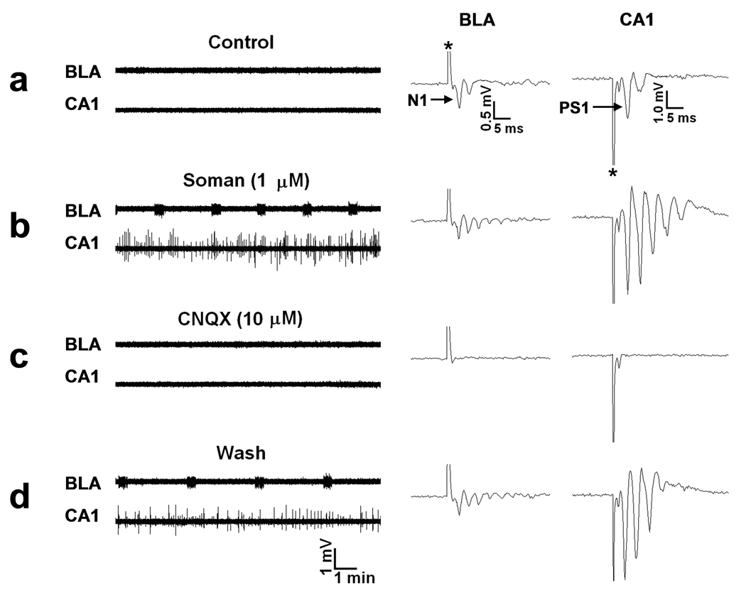
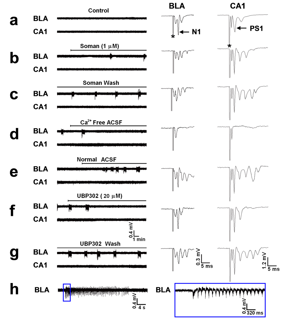
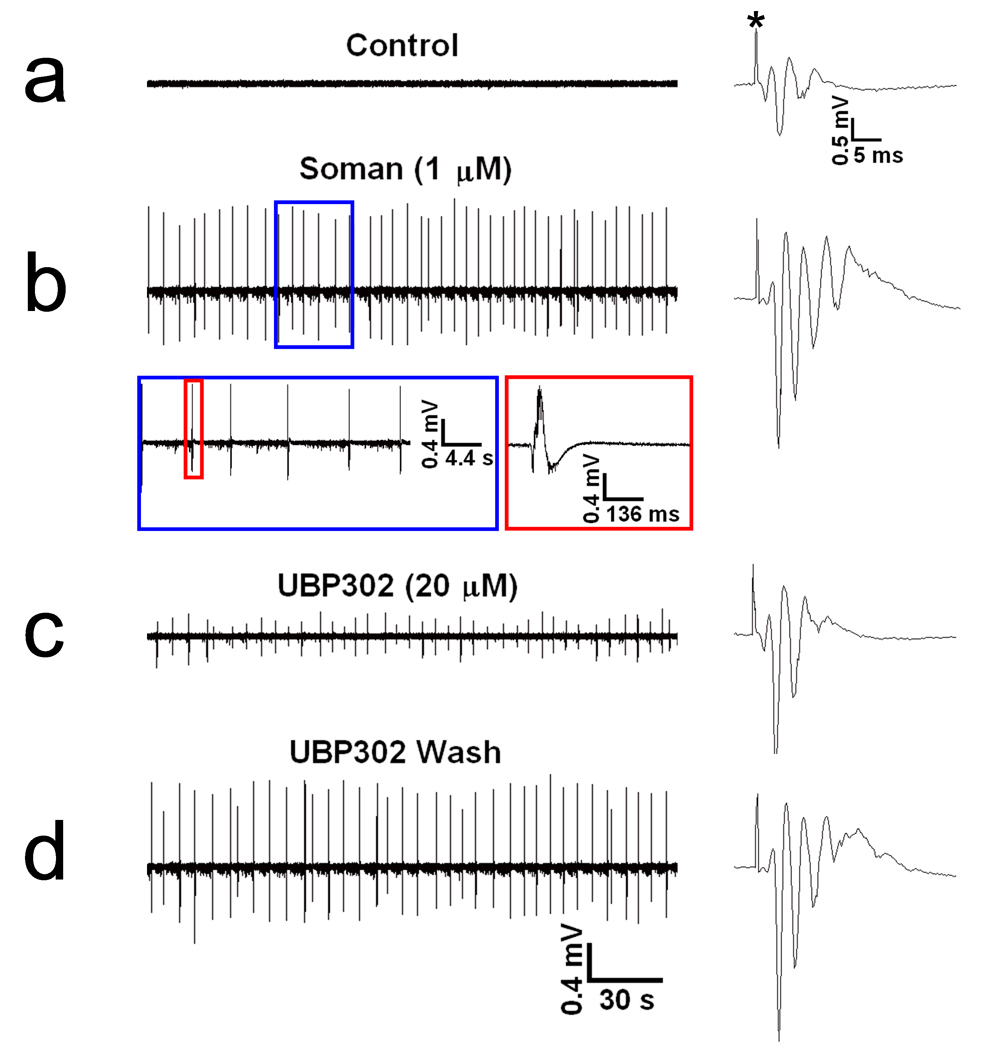
Exposure to organophosphorus nerve agents induces brain seizures, which can cause profound brain damage resulting in death or long-term cognitive deficits. The amygdala and the hippocampus are two of the most seizure-prone brain structures, but their relative contribution to the generation of seizures after nerve agent exposure is unclear. Here, we report that application of 1 muM soman for 30 min, in rat coronal brain slices containing both the hippocampus and the amygdala, produces prolonged synchronous neuronal discharges (10-40 s duration, 1.5-5 min interval of occurrence) resembling ictal activity in the basolateral nucleus of the amygdala (BLA), but only interictal-like activity ("spikes" of 100-250 ms duration; 2-5 s interval) in the pyramidal cell layer of the CA1 hippocampal area. BLA ictal- and CA1 interictal-like activity were synaptically driven, as they were blocked by the AMPA/kainate receptor antagonist 6-cyano-7-nitroquinoxaline-2,3-dione. As the expression of the GluR5 subunit of kainate receptors is high in the amygdala, and kainate receptors containing this subunit (GluR5KRs) play an important role in the regulation of neuronal excitability in both the amygdala and the hippocampus, we tested the efficacy of a GluR5KR antagonist against the epileptiform activity induced by soman. The GluR5KR antagonist UBP302 reduced the amplitude of the hippocampal interictal-like spikes, and eliminated the seizure-like discharges in the BLA, or reduced their duration and frequency, with no significant effect on the evoked field potentials. This is the first study reporting in vitro ictal-like activity in response to a nerve agent. Our findings, along with previous literature, suggest that the amygdala may play a more important role than the hippocampus in the generation of seizures following soman exposure, and provide the first evidence that GluR5KR antagonists may be an effective treatment against nerve agent-induced seizures.


Figures

FIG. 1
FIG. 1. Soman-induced ictal activity in the BLA and interictal activity in the hippocampus are synaptically driven
Extracellular field recordings, in gap-free mode, were simultaneously obtained in the BLA and the stratum pyramidale of the CA1 hippocampal area, in slices containing both regions. In this and the subsequent figures, the stimulus artifacts in the evoked field potentials (right panels) are indicated with an asterisk. (a) Field potentials in the BLA, evoked by stimulation of the external capsule, consisted of one major negative component (N1), followed by one or more lower-amplitude, late components. In the CA1 area, field potentials evoked by stimulation of the Schaffer collaterals consisted of a large population spike (PS1), which was often followed by one or two smaller amplitude, negative components. No spontaneous activity was present in the BLA or the CA1 area. (b) Exposure to 1 µM soman for 30 min reduced the amplitude of N1 in the BLA, and induced spontaneous, prolonged episodes of synchronous neuronal discharges resembling brain seizures. In response to soman exposure, the CA1 area produced additional population spikes in the enhanced evoked response, as well as spontaneous, interictal-like bursts. (c) Bath application of 10 µM CNQX (an AMPA/kainite receptor antagonist) blocked all synaptically-evoked components of the field potentials, as well as the BLA seizures and the CA1 interictal spikes. (d) The effects of CNQX were reversible.
FIG. 2
FIG. 2. Soman-induced ictal activity in the BLA is blocked by a GluR5KR antagonist
Extracellular field recordings were simultaneously obtained in the BLA and the stratum pyramidale of the CA1 hippocampal area, in slices containing both regions. (a) There was no spontaneous activity during control recordings in either the BLA or the CA1 area. N1 and PS1 of the BLA and CA1 field potentials, evoked by external capsule and Schaffer collateral pathway stimulation, respectively, were each followed by a smaller amplitude negative component, while a short latency fast component (non-synaptic; see d) was relatively pronounced in both regions, in this slice. (b) Exposure to 1 µM soman reduced the amplitude of N1 in the BLA, and produced seizure-like activity within 13 min of soman exposure. The first 3 seizures appeared at a 6 min interval, which was soon reduced to 5 min and then 4 min. Additional late negative components appeared in both the BLA and CA1 evoked field potentials, but no spontaneous activity was induced in the hippocampus in this slice. (c) The BLA seizures were not affected after soman washout. (d) Perfusion with Ca++ -free ACSF blocked both the BLA seizures and the synaptically-evoked components of the field potentials, and revealed the non-synaptic nature (fiber volley or antidromic) of the earliest, short-latency components of the evoked responses. (e) The BLA seizures and the evoked field responses returned upon reperfusion with normal medium (2 mM Ca++). (f) Bath application of 20 µM UBP302 (a GluR5KR antagonist) blocked the BLA seizures without affecting significantly the evoked field potentials, except for a reduction in the late components. (g) The effects of UBP302 were reversible. (h) A BLA seizure on an expanded time base. Oscillations had a higher frequency and amplitude at the beginning of the seizure, and dissipated gradually over the course of the seizure. The section of the trace within the blue rectangle is shown on the right with the time base further expanded.
FIG. 3
FIG. 3. Soman-induced interictal spikes in the hippocampus are reduced by a GluR5KR antagonist
The extracellular field recordings presented here are from the pyramidal cell layer of the CA1 hippocampal area; in this experiment the BLA did not produce spontaneous activity in response to soman and, for this reason, it is not shown. (a) In control conditions there was no spontaneous activity, while the evoked field potential consisted of PS1 and a second, smaller population spike. (b) Exposure to 1 µM soman for 30 min increased the number of population spikes in the evoked field potential and induced spontaneous interictal-like spikes, within 7 min of exposure; these spikes were unaffected by soman washout. The section of trace b within the blue rectangle is shown with an expanded time base, and the spike within the red rectangle is shown on the right with the time base further expanded. (c) Bath application of 20 µM UBP302 reduced the amplitude of the spontaneous spikes, without affecting their frequency. (d) The effects of UBP302 were reversible.

References

    1. Abou-Donia MB. Organophosphorus ester-induced delayed neurotoxicity. Annu Rev Pharmacol Toxicol. 1981;21:511–548. - PubMed
    1. Apland J. Effects of anticonvulsant drugs on soman-induced epileptiform activity in guinea pig hippocampal-entorhinal slices. USAMRICD-TR-01-02. Aberdeen Proving Ground, MD: U.S. Army Medical Research Institute of Chemical Defense; 2001.
    1. Aroniadou-Anderjaska V, Post RM, Rogawski MA, Li H. Input-specific LTP and depotentiation in the basolateral amygdala. NeuroReport. 2001;2:635–640. - PubMed
    1. Aroniadou-Anderjaska V, Qashu F, Braga MF. Mechanisms regulating GABAergic inhibitory transmission in the basolateral amygdala: Implications for epilepsy and anxiety disorders. Amino Acids. 2007;32:305–315. - PubMed
    1. Aroniadou-Anderjaska V, Fritsch B, Qashu F, Braga MF. Pathology and pathophysiology of the amygdala in epileptogenesis and epilepsy. Epilepsy Res. 2008;78:102–116. - PMC - PubMed

Publication types

MeSH terms