Review
. 2009 Jan;22(1):76-98, Table of Contents.
doi: 10.1128/CMR.00034-08.

Immunobiology of human cytomegalovirus: from bench to bedside

Affiliations
Review

Immunobiology of human cytomegalovirus: from bench to bedside

Tania Crough et al. Clin Microbiol Rev. 2009 Jan.

Abstract

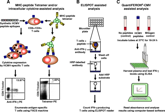
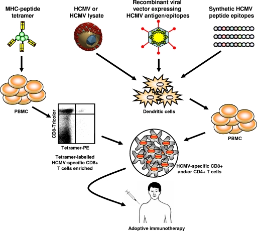
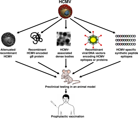
Following primary infection, human cytomegalovirus (HCMV) establishes lifelong latency and periodically reactivates without causing symptoms in healthy individuals. In the absence of an adequate host-derived immune response, this fine balance of permitting viral reactivation without causing pathogenesis is disrupted, and HCMV can subsequently cause invasive disease and an array of damaging indirect immunological effects. Over the last decade, our knowledge of the immune response to HCMV infection in healthy virus carriers and diseased individuals has allowed us to translate these findings to develop better diagnostic tools and therapeutic strategies. The application of these emerging technologies in the clinical setting is likely to provide opportunities for better management of patients with HCMV-associated diseases.


Figures

FIG. 1.
FIG. 1.
Comparative schematic genome organizations of human herpesviruses. VZV, varicella zoster virus; HSV, human simplex virus; EBV, Epstein-Barr virus. The lettering within the individual regions of the genome depicts the following features: terminal repeat long (TRL), unique long (UL), unique short (US), internal repeat long (IRL), internal repeat short (IRS), terminal repeat short (TRS), and internal repeat (IR).
FIG. 2.
FIG. 2.
Virtual three-dimensional model of HCMV showing various components of the virus. (Adapted from http://www.biografix.de/ with permission.)
FIG. 3.
FIG. 3.
Life cycle of HCMV in a human cell. HCMV enters human cells either through direct fusion or through the endocytic pathway. The virus attaches to the cell via interactions between viral glycoproteins (e.g., gB and gH) and a specific surface receptor(s) (e.g., platelet-derived growth factor α), followed by the fusion of the envelope with the cellular membrane to release nucleocapsids into the cytoplasm. These nucleocapsids are translocated into the nucleus, where viral DNA is released. This initiates the expression of IE-1/IE-2 genes. Viral replication and maturation follow the stimulation and parallel accumulation of viral synthesis function. This process involves the encapsulation of replicated viral DNA as capsids, which are then transported from the nucleus to the cytoplasm. Secondary envelopment occurs in the cytoplasm at the endoplasmic reticulum (ER)-Golgi intermediate compartment. This is followed by a complex two-stage final envelopment and egress process that leads to virion release by exocytosis at the plasma membrane.
FIG. 4.
FIG. 4.
Immune control of HCMV by innate and adaptive immunity. Primary infection with HCMV in healthy individuals typically initiates with replication in mucosal epithelium (A), after which the virus disseminates to monocytic cells of myeloid lineage including monocytes and CD34+ cells, where it establishes latent infection (B). Restricted viral gene expression is observed in these latently infected cells, thus limiting their immune recognition by effector cells. The differentiation of these virus-infected monocytes into macrophages can initiate productive infection (C). Virus particles or virus-associated dense bodies can be processed by professional antigen-presenting cells (e.g., DCs), which can stimulate antigen-specific T cells (D). In addition, these DCs activated through TLRs can also secrete a range of cytokines/chemokines, which activate the innate arm of the immune system (e.g., NK cells) (D). Virus-infected macrophages can also directly stimulate antigen-specific T cells (C). These activated T cells (CD8+, CD4+, and/or γδ T cells) and NK cells can directly lyse virus-infected cells by cytolysis or block virus replication through the secretion of cytokines such as IFN-γ and/or TNF (E). Another important arm of adaptive immunity involves B cells, which are also activated by the professional antigen-presenting cells and control extracellular virus through antibody-mediated neutralization (F).
FIG. 5.
FIG. 5.
Distribution of CD4+ and CD8+ T-cell responses within HCMV-encoded proteins. (A) Relative strengths of T-cell responses directed toward HCMV-encoded proteins with respect to expression kinetics (left) or gene function (right). (B) Schematic representation of the magnitude of CD4+ and CD8+ T-cell responses against immunodominant HCMV-encoded ORFs. The data presented in this figure are collated from data reported previously (69, 123-127, 133, 164, 263).
FIG. 6.
FIG. 6.
Ex vivo monitoring of HCMV-specific T-cell responses. (A) Peripheral blood mononuclear cells or whole blood is incubated with either an MHC-peptide tetramer or synthetic peptide epitopes. Following incubation, these cells are processed for flow cytometric analysis for the detection of antigen-specific T cells. For intracellular cytokine staining assays, the T cells were costained with anti-CD3, anti-CD4, anti-CD8, and anti-IFN-γ. For MHC-peptide tetramer analysis, the cells were stained with the MHC-peptide tetramer and anti-CD3, anti-CD4, and anti-CD8. Both these assays can be used to phenotypically characterize antigen-specific T cells using a variety of surface markers (see the text). PE, phycoerythrin; FACS, fluorescence-activated cell sorter. (B) For ELISPOT assays, peripheral blood mononuclear cells were stimulated with synthetic peptide, and IFN-γ was then captured using specific antibodies. This IFN-γ was detected using horseradish peroxidase (HRP)-labeled antibodies, and specific spot-forming cells were analyzed using image analysis software. (C) For the QuantiFERON-CMV assay, whole blood was stimulated with pooled HCMV peptide epitopes or mitogen, and IFN-γ in the plasma was detected and quantitated using standard enzyme-linked immunosorbent assay (ELISA) methodologies. (Panel C courtesy of Cellestis R&D Pty., Ltd.)
FIG. 7.
FIG. 7.
Strategies for adoptive immunotherapy of HCMV. A number of strategies have been explored for adoptive immunotherapy of HCMV. These include MHC-peptide tetramer enrichment of HCMV-specific T cells or in vitro stimulation of T cells with HCMV viral lysate, recombinant viral vectors, or synthetic peptides. Following enrichment or in vitro expansion, these cells are adoptively transferred into immunocompromised individuals either as a prophylactic or therapeutic treatment. These strategies have been reported previously (47, 66, 173, 194, 216, 217, 276). PE, phycoerythrin; PBMC, peripheral blood mononuclear cells.
FIG. 8.
FIG. 8.
Human cytomegalovirus vaccine development strategies. Various vaccine strategies have been developed, including live attenuated, recombinant viral proteins, dense bodies, vector vaccine subunit, or synthetic peptide epitopes. These formulations have been extensively tested using different animal models and have shown promising immunogenicity and protective efficacy (21, 30, 100, 228-230). Some of these strategies have already progressed to clinical trials with humans (1, 3, 18, 67, 75, 88, 104, 118, 176, 182, 191, 198, 199).

References

    1. Adler, S. P., S. H. Hempfling, S. E. Starr, S. A. Plotkin, and S. Riddell. 1998. Safety and immunogenicity of the Towne strain cytomegalovirus vaccine. Pediatr. Infect. Dis. J. 17:200-206. - PubMed
    1. Adler, S. P., G. Nigro, and L. Pereira. 2007. Recent advances in the prevention and treatment of congenital cytomegalovirus infections. Semin. Perinatol. 31:10-18. - PubMed
    1. Adler, S. P., S. A. Plotkin, E. Gonczol, M. Cadoz, C. Meric, J. B. Wang, P. Dellamonica, A. M. Best, J. Zahradnik, S. Pincus, K. Berencsi, W. I. Cox, and Z. Gyulai. 1999. A canarypox vector expressing cytomegalovirus (CMV) glycoprotein B primes for antibody responses to a live attenuated CMV vaccine (Towne). J. Infect. Dis. 180:843-846. - PubMed
    1. Ahn, K., A. Gruhler, B. Galocha, T. R. Jones, E. J. Wiertz, H. L. Ploegh, P. A. Peterson, Y. Yang, and K. Fruh. 1997. The ER-luminal domain of the HCMV glycoprotein US6 inhibits peptide translocation by TAP. Immunity 6:613-621. - PubMed
    1. Akbar, A. N., and J. M. Fletcher. 2005. Memory T cell homeostasis and senescence during aging. Curr. Opin. Immunol. 17:480-485. - PubMed

MeSH terms