

Epidemiology, diagnosis, treatment, and control of trichinellosis
- PMID: 19136437
- PMCID: PMC2620635
- DOI: 10.1128/CMR.00026-08
Epidemiology, diagnosis, treatment, and control of trichinellosis
Abstract
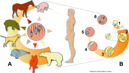
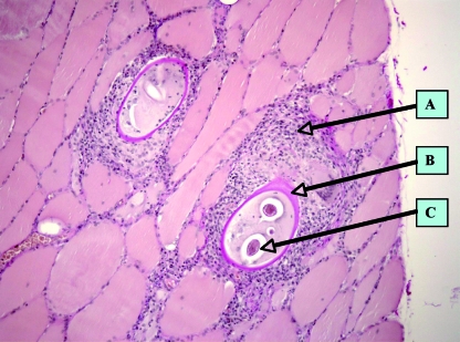
Throughout much of the world, Trichinella spp. are found to be the causative agents of human trichinellosis, a disease that not only is a public health hazard by affecting human patients but also represents an economic problem in porcine animal production and food safety. Due to the predominantly zoonotic importance of infection, the main efforts in many countries have focused on the control of Trichinella or the elimination of Trichinella from the food chain. The most important source of human infection worldwide is the domestic pig, but, e.g., in Europe, meats of horses and wild boars have played a significant role during outbreaks within the past 3 decades. Infection of humans occurs with the ingestion of Trichinella larvae that are encysted in muscle tissue of domestic or wild animal meat. Early clinical diagnosis of trichinellosis is rather difficult because pathognomonic signs or symptoms are lacking. Subsequent chronic forms of the disease are not easy to diagnose, irrespective of parameters including clinical findings, laboratory findings (nonspecific laboratory parameters such as eosinophilia, muscle enzymes, and serology), and epidemiological investigations. New regulations laying down rules for official controls for Trichinella in meat in order to improve food safety for consumers have recently been released in Europe. The evidence that the disease can be monitored and to some extent controlled with a rigorous reporting and testing system in place should be motivation to expand appropriate programs worldwide.
Figures
References
-
- Akkoc, N., Z. Kuruuzum, S. Akar, A. Yuce, F. Onen, N. Yapar, O. Ozgenc, M. Turk, D. Ozdemir, M. Avci, Y. Guruz, A. M. Oral, and E. Pozio. 2008. A large scale outbreak of trichinellosis due to Trichinella britovi in Turkey. Zoonoses Public Health. doi: 10.1111/j.1863-2378.2008.01158.x. - DOI - PubMed
-
- Ancelle, T., A. E. De Bruyne, and J. Dupouy-Camet. 2005. Outbreak of trichinellosis due to consumption of bear meat from Canada, France, September 2005. Euro Surveill. 10:EO51013.3. - PubMed
-
- Ancelle, T. 1998. History of trichinellosis outbreaks linked to horse meat consumption 1975-1998. Euro Surveill. 3:86-89. - PubMed
-
- Anonymous. 2004. Drugs for parasitic infections. Med. Lett. 2004:1-12.
-
- Appleton, J. A., R. G. Bell, W. Homan, and F. Van Knapen. 1991. Consensus on Trichinella spiralis antigens and antibodies. Parasitol. Today 7:190-192.
Publication types
MeSH terms
LinkOut - more resources
Full Text Sources
Other Literature Sources
Medical
Miscellaneous
