. 2009 Mar;8(3):410-20.
doi: 10.1128/EC.00256-08. Epub 2009 Jan 9.

The G-alpha protein GNA3 of Hypocrea jecorina (Anamorph Trichoderma reesei) regulates cellulase gene expression in the presence of light

Affiliations

The G-alpha protein GNA3 of Hypocrea jecorina (Anamorph Trichoderma reesei) regulates cellulase gene expression in the presence of light

Monika Schmoll et al. Eukaryot Cell. 2009 Mar.

Abstract

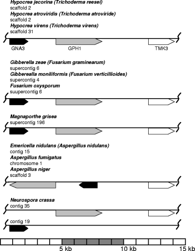
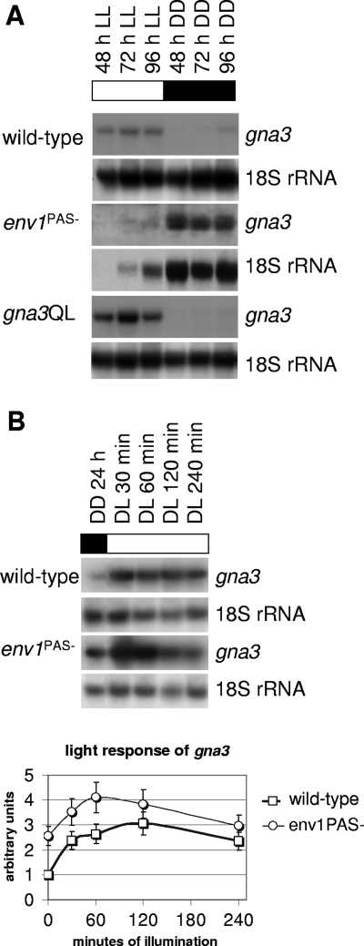
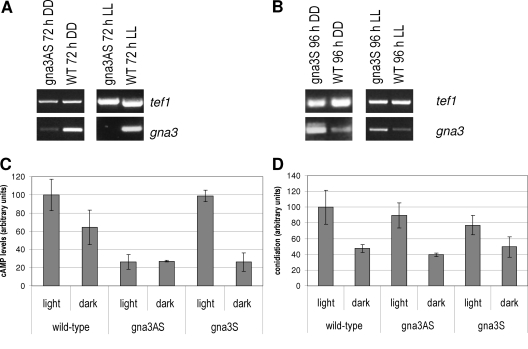
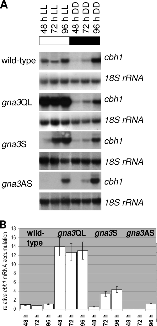
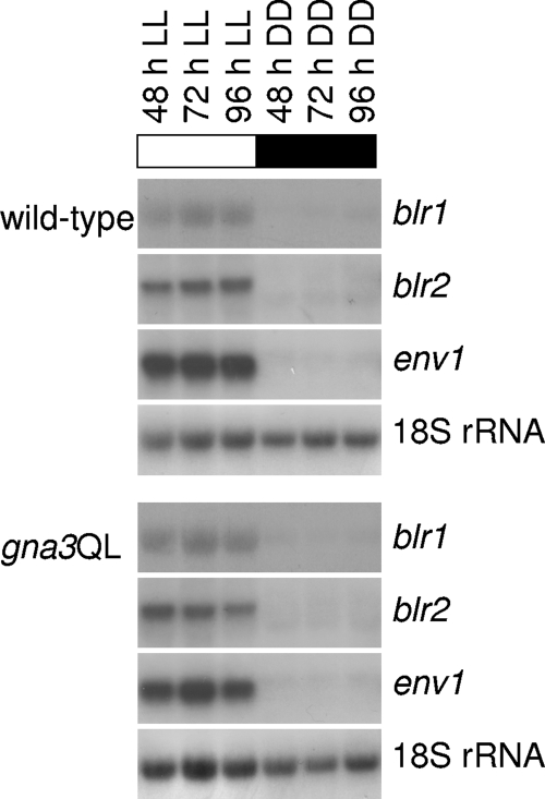
Although the enzymes enabling Hypocrea jecorina (anamorph Trichoderma reesei) to degrade the insoluble substrate cellulose have been investigated in some detail, little is still known about the mechanism by which cellulose signals its presence to the fungus. In order to investigate the possible role of a G-protein/cyclic AMP signaling pathway, the gene encoding GNA3, which belongs to the adenylate cyclase-activating class III of G-alpha subunits, was cloned. gna3 is clustered in tandem with the mitogen-activated protein kinase gene tmk3 and the glycogen phosphorylase gene gph1. The gna3 transcript is upregulated in the presence of light and is almost absent in the dark. A strain bearing a constitutively activated version of GNA3 (gna3QL) exhibits strongly increased cellulase transcription in the presence of the inducer cellulose and in the presence of light, whereas a gna3 antisense strain showed delayed cellulase transcription under this condition. However, the gna3QL mutant strain was unable to form cellulases in the absence of cellulose. The necessity of light for stimulation of cellulase transcription by GNA3 could not be overcome in a mutant which expressed gna3 under control of the constitutive gpd1 promoter also in darkness. We conclude that the previously reported stimulation of cellulase gene transcription by light, but not the direct transmission of the cellulose signal, involves the function and activation of GNA3. The upregulation of gna3 by light is influenced by the light modulator ENVOY, but GNA3 itself has no effect on transcription of the light regulator genes blr1, blr2, and env1. Our data for the first time imply an involvement of a G-alpha subunit in a light-dependent signaling event in fungi.


Figures

FIG. 1.
FIG. 1.
Gene structure of gna3, transformation with the gna3Q206L allele, and PCR strategy. (A) The gene structure of gna3 is shown along with its exons and introns, the predicted GNA3 protein, and construction of a transformation cassette to introduce the single amino acid exchange at position 206 as indicated by a triangle. The EcoRI and XbaI restriction sites have been introduced using nested primers to facilitate cloning. (B) Southern blot analysis of TU-6 and two Q206L mutants. Genomic DNA was digested with HindIII or EcoRI/XbaI and probed with an ∼3.2-kb EcoRI/XbaI fragment excised from pBgna3Q206L. The additional HindIII band at 1.6 kb results from one restriction site within gna3 and a second site within the vector backbone, and the bands around 10 kb result from ectopic integration of the cassette and indicate the presence of only one copy in either of the two strains used for further experiments. The second Southern blot resulting from digestion with EcoRI/XbaI confirms ectopic integration of the mutant allele, since the 8,843-bp wild-type fragment is present in all three strains. Only the mutant strains contain the 3.2-kb fragment resulting from the artificially introduced restriction sites.
FIG. 2.
FIG. 2.
Composition of the MGG cluster in ascomycetes. Contig or scaffold numbers are represented as specified in the respective genome databases (http://www.broad.mit.edu/annotation/fungi/; http://genome.jgi-psf.org/). Genes encoding orthologues of GNA3 are given in black, those encoding orthologues of GPH1 are in gray, and those encoding orthologues of TMK3 are in white. The scheme is drawn to scale. For exact genomic coordinates and known orthologues of the respective genes, see Table S1 in the supplemental material.
FIG. 3.
FIG. 3.
gna3 transcription is enhanced by light. (A) Mycelia of the wild-type strain TU-6 and of the env1PAS− and gna3QL mutant strains were cultivated in Mandels-Andreotti medium with 1% (wt/vol) microcrystalline cellulose as the sole carbon source in constant darkness (DD) or constant light (LL). Twenty micrograms of total RNA was loaded per lane, and an α-32P-radiolabeled PCR fragment spanning the cDNA of gna3 was used as a probe. 18S rRNA was used as control. (B) The short-term light response of gna3 was analyzed in the wild-type strain QM9414 and the env1PAS− mutant strain upon growth in Mandels-Andreotti medium with 1% (wt/vol) glycerol as a carbon source in constant darkness (DD) and after 30, 60, 120, and 240 min of illumination (DL). Twenty micrograms of total RNA was loaded per lane, and an α-32P-radiolabeled PCR fragment spanning the cDNA of gna3 was used as a probe. Results were quantified, and the amount of light-induced transcription of gna3 in the specified strain was normalized to the 18S rRNA control hybridization. Graphs show transcription levels above background.
FIG. 4.
FIG. 4.
RT-PCR and cAMP levels of gna3 antisense and overexpressing strains. (A) RT-PCR of gna3 in the gna3AS antisense strain after 72 h of growth in Mandels-Andreotti medium with cellulose as a carbon source in constant light (LL) and constant darkness (DD). tef1 (encoding translation elongation factor 1 alpha) was used as a control. The antisense fragment of gna3 is under the control of the constitutive gpd1 promoter in the gna3AS strain. (B) RT-PCR of gna3 in the gna3S overexpressing strain under conditions where transcription of gna3 was below the detection limit in Northern blots, i.e., after 96 h of growth in the same medium as mentioned above in constant darkness (DD) and additionally after 96 h in constant light (LL). gna3 is under the control of the constitutive gpd1 promoter in the gna3S strain. (C) cAMP levels in the gna3AS and gna3S strains after 72 h of growth in constant light or constant darkness on Mandels-Andreotti medium with cellulose as a carbon source relative to the wild-type strain (21.72 pmol/mg protein). (D) Conidiation of the gna3AS and gna3S strains relative to the wild type in light and darkness.
FIG. 5.
FIG. 5.
Northern analysis of cellulase gene transcription and gna3. (A) Mycelia of the wild-type (TU-6), gna3QL (constitutively activated GNA3, expression under the control of the original gna3 promoter), gna3AS (antisense strain, fragment under the control of the constitutive gpd1 promoter), and gna3S (overexpressing strain, gna3 under the control of the gpd1 promoter) strains were grown in Mandels-Andreotti medium supplemented with 1% microcrystalline cellulose as a carbon source in constant light (1,800 lx, 25 μmol photons m−2 s−1) (LL) or constant darkness (DD) for 48, 72, and 96 h. For Northern blotting, 20 μg of total RNA was loaded per lane and α-32P-radiolabeled PCR fragments of cbh1 and 18S rRNA (control) were used as probes. (B) Quantitative analysis of transcript abundance in constant light on cellulose in the wild-type, gna3QL, gna3AS, and gna3S strains. Values were normalized to wild type and to the corresponding 18S rRNA using the Bio-Rad GS-800 calibrated densitometer.
FIG. 6.
FIG. 6.
GNA3 does not influence transcription of the light receptors blr1, blr2, and env1. Mycelia of the wild-type (TU-6) and gna3QL strains were grown in Mandels-Andreotti medium supplemented with 1% (wt/vol) microcrystalline cellulose as a carbon source in constant light (1,800 lx, 25 μmol photons m−2 s−1) (LL) for 48, 72, and 96 h or in constant darkness (DD). For Northern blotting, 20 μg of total RNA was loaded per lane and α-32P-radiolabeled PCR fragments of blr1, blr2, env1, and 18S rRNA were used as probes.

References

    1. Amaar, Y. G., and M. M. Moore. 1998. Mapping of the nitrate-assimilation gene cluster (crnA-niiA-niaD) and characterization of the nitrite reductase gene (niiA) in the opportunistic fungal pathogen Aspergillus fumigatus. Curr. Genet. 33206-215. - PubMed
    1. Aro, N., T. Pakula, and M. Penttila. 2005. Transcriptional regulation of plant cell wall degradation by filamentous fungi. FEMS Microbiol. Rev. 29719-739. - PubMed
    1. Aro, N., A. Saloheimo, M. Ilmen, and M. Penttila. 2001. ACEII, a novel transcriptional activator involved in regulation of cellulase and xylanase genes of Trichoderma reesei. J. Biol. Chem. 27624309-24314. - PubMed
    1. Baxevanis, A. D., and D. Landsman. 1995. The HMG-1 box protein family: classification and functional relationships. Nucleic Acids Res. 231604-1613. - PMC - PubMed
    1. Berman, D. M., T. M. Wilkie, and A. G. Gilman. 1996. GAIP and RGS4 are GTPase-activating proteins for the Gi subfamily of G protein alpha subunits. Cell 86445-452. - PubMed

Publication types

MeSH terms

Associated data

LinkOut - more resources