. 2009 Mar;296(3):H627-38.
doi: 10.1152/ajpheart.00444.2008. Epub 2009 Jan 9.

Cardiac fibroblasts require focal adhesion kinase for normal proliferation and migration

Affiliations

Cardiac fibroblasts require focal adhesion kinase for normal proliferation and migration

Ana Maria Manso et al. Am J Physiol Heart Circ Physiol. 2009 Mar.

Abstract

Migration and proliferation of cardiac fibroblasts (CFs) play an important role in the myocardial remodeling process. While many factors have been identified that regulate CF growth and migration, less is known about the signaling mechanisms involved in these processes. Here, we utilized Cre-LoxP technology to obtain focal adhesion kinase (FAK)-deficient adult mouse CFs and studied how FAK functioned in modulating cell adhesion, proliferation, and migration of these cells. Treatment of FAK(flox/flox) CFs with Ad/Cre virus caused over 70% reduction of FAK protein levels within a cell population. FAK-deficient CFs showed no changes in focal adhesions, cell morphology, or protein expression levels of vinculin, talin, or paxillin; proline-rich tyrosine kinase 2 (Pyk2) expression and activity were increased. Knockdown of FAK protein in CFs increased PDGF-BB-induced proliferation, while it reduced PDGF-BB-induced migration. Adhesion to fibronectin was not altered. To distinguish between the function of FAK and Pyk2, FAK function was inhibited via adenoviral-mediated overexpression of the natural FAK inhibitor FAK-related nonkinase (FRNK). Ad/FRNK had no effect on Pyk2 expression, inhibited the PDGF-BB-induced migration, but did not change the PDGF-BB-induced proliferation. FAK deficiency had only modest effects on increasing PDGF-BB activation of p38 and JNK MAPKs, with no alteration in the ERK response vs. control cells. These results demonstrate that FAK is required for the PDGF-BB-induced migratory response of adult mouse CFs and suggest that FAK could play an essential role in the wound-healing response that occurs in numerous cardiac pathologies.


Figures

Fig. 1.
Fig. 1.
Excision of the focal adhesion kinase (FAK) gene in FAKflox/flox cardiac fibroblasts (CFs) by recombinant adenoviral Cre-recombinase virus (Ad/Cre). A: diagrams of FAK floxed and excised alleles. Primers used for identification of the unexcised and excised alleles are P1 and P2 (note: diagrams are not drawn to scale). B: PCR fragments from intact or excised floxed alleles were detected at 1.6 kb or 350 bp, respectively. The 350-bp excised fragment was detected in FAKflox/flox CFs infected with Ad/Cre virus at both 100 and 200 multiplicity of infection (MOI). No excised fragment was detected in FAKflox/flox CFs infected with adenovirus expressing β-galactosidase (Ad/LacZ) (control) virus.
Fig. 2.
Fig. 2.
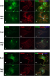
Localization of FAK, proline-rich tyrosine kinase 2 (Pyk2), and vinculin (Vin) in control (Cntl) and FAK-deficient CFs. FAK (green; A and D), actin (red; B and E), and 4,6-diamidino-2-phenylindole (DAPI) (blue; C and F) are shown in Cntl (AC) and FAK knockout (KO) cells (DF). PYK2 (green; G and J), actin (red; H and K), and DAPI (blue; I and L) are shown in Cntl (GI) and FAK KO cells (JL). Vin (green; M and P), actin (red; N and Q), and DAPI (blue; O and R) are shown in Cntl (MO) and FAK KO cells (PR). Immunofluorescent staining of FAKflox/flox CFs infected with Ad/Cre (FAK KO) or Ad/LacZ (Cntl) showed that FAK protein was localized in focal adhesions (FAs) and nucleus in Cntl cells (A and C). The amount of FAK was reduced toward background levels in Ad/Cre-infected cells compared with Ad/LacZ-infected cells (D and F). Pyk2 showed a similar distribution to FAK, localizing in FAs and nucleus in Cntl cells (G and I), and no changes in Pyk2 localization were seen between Cntl and FAK KO cells (G and I; J and L). Vin, a marker of FAs, showed a similar distribution in both Cntl and FAK KO cells (M and O; P and R). Phalloidin (red) was used to mark F-actin, and DAPI (blue) labeled nuclei.
Fig. 3.
Fig. 3.
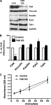
Reduction of FAK expression alters expression of FA proteins but does not change adhesion to fibronectin of adult CFs. A: lysates from FAKflox/flox CFs infected with Ad/Cre or Ad/LacZ (200 MOI) for 96 h were analyzed by Western blotting for FAK and other key FA proteins: Vin, paxillin, Pyk2, and talin. GAPDH was used as a loading Cntl. Blots are representative of 5 independent experiments. B: densitometric analysis was performed on Western blots by normalizing FAK, Vin, talin, paxillin, and Pyk2 expression to simultaneously measured GAPDH. CFs infected with Ad/Cre showed a 70% reduction of FAK protein expression (**P < 0.001, n = 5) and 52% increase of Pyk2 (*P < 0.05, n = 5) compared with Cntl infected CFs. No other significant alterations in protein expression were found. Data represent mean values ± SE. C: adhesion of FAK-deficient CFs (Ad/Cre) to fibronectin was not different from that of Cntl cells when evaluated at 10, 30, and 60 min following plating on a fibronectin matrix.
Fig. 4.
Fig. 4.
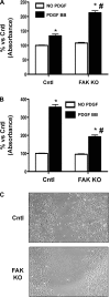
FAK-deficient CFs have reduced migration and increased proliferation in response to platelet-derived growth factor (PDGF)-BB. A: cell proliferation was increased in FAK-deficient CFs, as determined by 5-bromo-2′-deoxyuridine (BrdU) incorporation. FAK KO or Cntl CFs were incubated for 24 h with PDGF-BB (10 ng/ml), with the last 2 h in the presence of BrdU. FAK KO CFs showed an increase in proliferation compared with Cntl CFs (#P < 0.001 vs. Cntl + PDGF BB; *P < 0.001 vs. No PDGF BB, n = 5). B: migration of FAK-deficient CFs was reduced as quantified using a Boyden chamber migration assay. FAK KO CFs had reduced migration toward 10 ng/ml PDGF-BB in a Boyden chamber assay compared with Cntl CFs when evaluated over 3 h (#P < 0.001 vs. Ad/LacZ + PDGF-BB; *P < 0.001 vs. No PDGF-BB; n = 3). Data represent mean values ± SE. C: wound assays show reduced migration of FAK KO CFs compared with Cntl cells when stimulated with 20 ng/ml of PDGF-BB. Matched pairs were photographed 48 h following PDGF stimulation.
Fig. 5.
Fig. 5.
FAK-related nonkinase (FRNK) expression inhibits PDGF-BB-stimulated migration in CFs but does not change proliferation. A and B: lysates from CFs infected with Ad/FRNK or Ad/LacZ (Cntl) (25 MOI, each) for 48 h were analyzed by Western blotting (A) with antibodies to FAK, Pyk2, and tubulin. The blots are representative of 3 independent experiments. B: densitometric analysis was performed by normalizing FAK and Pyk2 expression to simultaneously measured tubulin. Data represent mean values ± SE. C: cell proliferation of CFs was not altered by FRNK as determined by BrdU incorporation. CFs infected with Ad/FRNK or Ad/LacZ (Cntl) were incubated for 24 h with 10 ng/ml PDGF-BB with the last 2 h in the presence of BrdU. CFs infected with Ad/FRNK did not show any difference in proliferation compared with Cntl CFs (*P < 0.001 vs. No PDGF-BB, n = 4). D: FRNK causes reduced migration of CFs when quantified using a Boyden chamber migration assay. CFs infected with FRNK (Ad/FRNK) or LacZ (Cntl) were allowed to migrate for 3 h toward 10 ng/ml PDGF-BB. Increased expression of FRNK in CFs caused a reduction in migration compared with Cntl CFs (#P < 0.01 vs. Cntl + PDGF-BB, *P < 0.01 vs. No PDGF BB, +P < 0.05 vs. Cntl, n = 3). Data represent mean values ± SE.
Fig. 6.
Fig. 6.
FAK and Pyk2 activation in response to PDGF-BB. FAK KO or Cntl CFs were treated with 10 ng/ml PDGF-BB for various times as indicated. A: total and Tyr 397 phosphorylated FAK (p-FAK) protein were analyzed by Western blotting and showed no increase in phosphorylation in Cntl cells after PDGF-BB treatment. B: densitometric analysis was performed on Western blots by normalizing p-FAK (Tyr 397) to total FAK (n = 3). Data represent mean values ± SE. C: total and Tyr 402 phosphorylated Pyk2 (p-Pyk2) protein were analyzed by Western blotting and showed an increase in phosphorylation at Tyr 402 in Cntl and FAK KO cells in response to PDGF-BB. D: densitometric analysis was performed on Western blots by normalizing p-Pyk2 (Tyr 402) to total Pyk2 [*P < 0.05 vs. Cntl (No PDGF-BB), +P < 0.01 vs. FAK KO (No PDGF-BB), #P < 0.01 vs. Cntl+ PDGF-BB (30 min), n = 7]. Data represent mean values ± SE.
Fig. 7.
Fig. 7.
Pyk2 activation in response to PDGF-BB in FRNK-treated cells. Ad/FRNK or LacZ (Cntl) CFs were treated with 10 ng/ml PDGF-BB for 30 min. A: total and Tyr 402 p-Pyk2 protein were analyzed by Western blotting and showed no increase in phosphorylation in Ad/FRNK-treated cells after PDGF-BB treatment. B: densitometric analysis was performed on Western blots by normalizing p-Pyk2 (Tyr 402) to total Pyk2 (n = 5). *P < 0.05 vs. Cntl (No PDGF-BB). Data represent mean values ± SE.
Fig. 8.
Fig. 8.
Activation of MAPKs by PDGF-BB in FAK-deficient CFs. FAK KO or Cntl CFs were treated with 10 ng/ml PDGF-BB for various times as indicated. Activated and total ERK1/2 (A), JNK (B), and p-38 (C) were analyzed by Western blotting and showed only a trend toward increased phosphorylation of JNK and p38 in the FAK-deficient CFs compared with Cntl cells. Overall, no statistically significant differences in MAPK activation was noted in the KO cells. The blots are representative of at least three independent experiments.

Similar articles

Cited by

References

    1. Banerjee I, Fuseler JW, Price RL, Borg TK, Baudino TA. Determination of cell types and numbers during cardiac development in the neonatal and adult rat and mouse. Am J Physiol Heart Circ Physiol 293: H1883–H1891, 2007. - PubMed
    1. Baudino TA, Carver W, Giles W, Borg TK. Cardiac fibroblasts: friend or foe? Am J Physiol Heart Circ Physiol 291: H1015–H1026, 2006. - PubMed
    1. Beggs HE, Schahin-Reed D, Zang K, Goebbels S, Nave KA, Gorski J, Jones KR, Sretavan D, Reichardt LF. FAK deficiency in cells contributing to the basal lamina results in cortical abnormalities resembling congenital muscular dystrophies. Neuron 40: 501–514, 2003. - PMC - PubMed
    1. Braren R, Hu H, Kim YH, Beggs HE, Reichardt LF, Wang R. Endothelial FAK is essential for vascular network stability, cell survival, and lamellipodial formation. J Cell Biol 172: 151–162, 2006. - PMC - PubMed
    1. Brinson AE, Harding T, Diliberto PA, He Y, Li X, Hunter D, Herman B, Earp HS, Graves LM. Regulation of a calcium-dependent tyrosine kinase in vascular smooth muscle cells by angiotensin II and platelet-derived growth factor. Dependence on calcium and the actin cytoskeleton. J Biol Chem 273: 1711–1718, 1998. - PubMed

Publication types

MeSH terms

LinkOut - more resources