. 2009 Jan 1;23(1):93-104.
doi: 10.1101/gad.502409.

Second messenger-mediated spatiotemporal control of protein degradation regulates bacterial cell cycle progression

Affiliations

Second messenger-mediated spatiotemporal control of protein degradation regulates bacterial cell cycle progression

Anna Duerig et al. Genes Dev. .

Abstract

Second messengers control a wide range of important cellular functions in eukaryotes and prokaryotes. Here we show that cyclic di-GMP, a global bacterial second messenger, promotes cell cycle progression in Caulobacter crescentus by mediating the specific degradation of the replication initiation inhibitor CtrA. During the G1-to-S-phase transition, both CtrA and its cognate protease ClpXP dynamically localize to the old cell pole, where CtrA is rapidly degraded. Sequestration of CtrA to the cell pole depends on PopA, a newly identified cyclic di-GMP effector protein. PopA itself localizes to the cell pole and directs CtrA to this subcellular site via the direct interaction with a mediator protein, RcdA. We present evidence that c-di-GMP regulates CtrA degradation during the cell cycle by controlling the dynamic sequestration of the PopA recruitment factor to the cell pole. Furthermore, we show that cell cycle timing of CtrA degradation relies on converging pathways responsible for substrate and protease localization to the old cell pole. This is the first report that links cyclic di-GMP to protein dynamics and cell cycle control in bacteria.


Figures
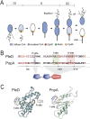
Figure 1.
Figure 1.
Dynamic protein localization and CtrA degradation during the cell cycle. (A) Schematic of CtrA, ClpXP, RcdA, and CpdR localization during the C. crescentus cell cycle. (B) Sequence alignment of the PleD and PopA. The amino acid sequence flanking the phosphoryl acceptor site (P-site), I-site, and A-site are shown with the conserved residues colored in red, the signature motifs boxed, and amino acids residues replaced in P-site, I-site, and A-site mutants marked with asterisks. (C) Comparison of the 3D structure of the GGDEF domains of PleD (as determined by X-ray crystallography) (Chan et al. 2004) and modeled PopA. A-sites and I-sites are marked, and the position of a dimer of c-di-GMP bound to the I-site is indicated.
Figure 2.
Figure 2.
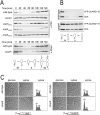
Cell cycle-dependent degradation of CtrA requires PopA. (A) Immunoblots of synchronized cultures of C. crescentus wild-type and popA mutant strains. Immunoblots were stained with anti-CtrA or anti-McpA antibodies as indicated. (B) Synchronized swarmer cells of wild type (top panel) and popA mutant (bottom panel) expressing yfp-ctrARD + 15 from the xylose-inducible promoter Pxyl were released into M2G minimal glucose medium and monitored throughout the cell cycle using anti-CtrA antibodies. The YFP-CtrARD + 15 fusion protein and wild-type CtrA are labeled. (C) Morphology and replication status of C. crescentus wild type and ΔpopA mutant expressing ctrA (left panels, strains UJ3966 and UJ3969) or ctrAD51E (right panels, strains UJ3967 and UJ3970) from the xylose-inducible promoter Pxyl. Cells were harvested under inducing (xylose) or non-inducing (glucose) conditions and analyzed by light microscopy and by flow cytometry.
Figure 3.
Figure 3.
PopA mediates polar localization of RcdA and CtrA. (A) Wild-type and ΔpopA mutant cells expressing yfp-ctrARD+15 were analyzed by DIC and fluorescence microscopy. Polar foci of Yfp-CtrARD + 15 are marked by arrows and shown schematically in the right panel. (B) Wild-type, ΔpopA, and popAR357G (I-site) mutant cells expressing rcdA-yfp were analyzed by DIC and fluorescence microscopy. (C) PopA directly interacts with RcdA. The red color on McConkey agar-base maltose plates is an indicator for protein–protein interaction. PopA and RcdA fusions to the adenylate cyclase fragments T18 and T25 are indicated. Zip indicates a positive control. (D) In vivo FRET analysis of PopA and RcdA. C. crescentus cells expressing rcdA-ecfp and popA-eyfp (left panel) or popA-ecfp and rcdA-eyfp (right panel) were analyzed. The intensity of the CFP channel was recorded before and after YFP-specific bleaching. The increase of signal intensity of the CFP channel after specific bleaching of the YFP channel is a measure of the FRET efficiency (%).
Figure 4.
Figure 4.
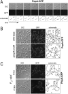
PopA localizes to the old and new cell poles. (A) Time-lapse experiment with synchronized C. crescentus wild-type cells expressing popA-egfp. The first sample corresponds to the 0-min time point, and samples were analyzed every 20 min. (B) RcdA is dispensable for PopA localization to the cell poles. C. crescentus wild-type and ΔrcdA mutant cells expressing popA-egfp were analyzed by DIC and fluorescence microscopy. (C) ClpX is dispensable for PopA localization to the cell poles. Mixed cultures of the C. crescentus conditional clpX mutant strain UJ271 expressing popA-egfp were analyzed by DIC and fluorescence microscopy under permissive (PYEX) and restrictive conditions (PYEG). Cultures were grown in the absence of xylose for 6 h, resulting in complete depletion of ClpX (data not shown).
Figure 5.
Figure 5.
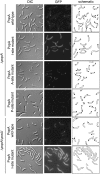
Distinct mechanisms mediate PopA localization to the new and old pole. C. crescentus wild type and podJ mutant expressing GFP fusion proteins to PopA wild type and the following PopA mutants were analyzed by DIC and fluorescence microscopy: PopAD55N (P-site mutant), PopAE368Q (A-site mutant), PopAR357G (I-site mutant). Polar localization is indicated schematically in the panels on the right.
Figure 6.
Figure 6.
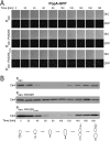
Expression of a heterologous phosphodiesterase interferes with PopA localization and CtrA degradation. Cell cycle-dependent PopA-GFP localization (A) and CtrA degradation (B) in cells of C. crescentus CB15 harboring a vanillate induced copy of PA5295 from P. aeruginosa (middle panels) or an active site mutant, PA5295E328A (bottom panels). Control assays with cells harboring the empty vector (Pvan) are shown in the top panel.
Figure 7.
Figure 7.
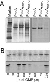
PopA binds c-di-GMP at the conserved I-site of the GGDEF domain. (A) UV cross-link experiment of purified hexahistidine-tagged PopA with [33P]-labeled c-di-GMP. The following proteins were used: PopA, PopAE368D (A-site mutant), PopAR357G (I-site mutant). The Coomassie blue-stained gel (left) and the autoradiograph (right) are shown. (B) UV cross-linking of purified PopA with [33P]-labeled c-di-GMP and increasing concentrations of nonlabeled c-di-GMP (0–80 μM). Coomassie blue-stained gel (top panel) and autoradiograph (bottom panel) are shown.
Figure 8.
Figure 8.
CpdR and PopA constitute two converging pathways leading to cell cycle-dependent degradation of CtrA. (A) Cultures of C. crescentus ΔcpdR single and ΔcpdRΔpopA double mutants expressing cpdRD51A-yfp under the control of the xylose-inducible promoter Pxyl were grown in the presence of xylose and analyzed microscopically. (B) Cultures of different C. crescentus ΔcpdR and ΔpopA mutants expressing cpdRD51A-yfp under the control of the xylose-inducible promoter Pxyl were grown in the presence (PYEX) or absence of xylose (PYEG) and analyzed by immunoblots using anti-CtrA antibodies.
Figure 9.
Figure 9.
Model for the role of PopA in cell cycle-dependent degradation of CtrA. (A) Converging pathways involved in polar sequestration of ClpXP and its substrate CtrA. The CckA-ChpT phosphorelay inversely regulates CtrA activity and stability through the phosphorylation of CtrA and CpdR. The model predicts that cell cycle-dependent localization of PopA to the stalked cell pole involves the timed synthesis and/or hydrolysis of c-di-GMP by as-yet-unidentified DGCs or PDEs. (B) Upon binding of c-di-GMP, PopA sequesters to the cell pole, where it recruits RcdA and CtrA.

References

    1. Alba B.M., Leeds J.A., Onufryk C., Lu C.Z., Gross C.A. DegS and YaeL participate sequentially in the cleavage of RseA to activate the σE-dependent extracytoplasmic stress response. Genes & Dev. 2002;16:2156–2168. - PMC - PubMed
    1. Aldridge P., Paul R., Goymer P., Rainey P., Jenal U. Role of the GGDEF regulator PleD in polar development of Caulobacter crescentus . Mol. Microbiol. 2003;47:1695–1708. - PubMed
    1. Altschul S.F., Madden T.L., Schaffer A.A., Zhang J., Zhang Z., Miller W., Lipman D.J. Gapped BLAST and PSI-BLAST: A new generation of protein database search programs. Nucleic Acids Res. 1997;25:3389–3402. - PMC - PubMed
    1. Berman H.M., Westbrook J., Feng Z., Gilliland G., Bhat T.N., Weissig H., Shindyalov I.N., Bourne P.E. The Protein Data Bank. Nucleic Acids Res. 2000;28:235–242. - PMC - PubMed
    1. Biondi E.G., Reisinger S.J., Skerker J.M., Arif M., Perchuk B.S., Ryan K.R., Laub M.T. Regulation of the bacterial cell cycle by an integrated genetic circuit. Nature. 2006;444:899–904. - PubMed

Publication types

MeSH terms

LinkOut - more resources