. 2009 Jan 27;106(4):1117-21.
doi: 10.1073/pnas.0809144106. Epub 2009 Jan 9.

Insulin levels control female germline stem cell maintenance via the niche in Drosophila

Affiliations

Insulin levels control female germline stem cell maintenance via the niche in Drosophila

Hwei-Jan Hsu et al. Proc Natl Acad Sci U S A. .

Abstract

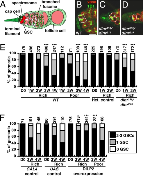
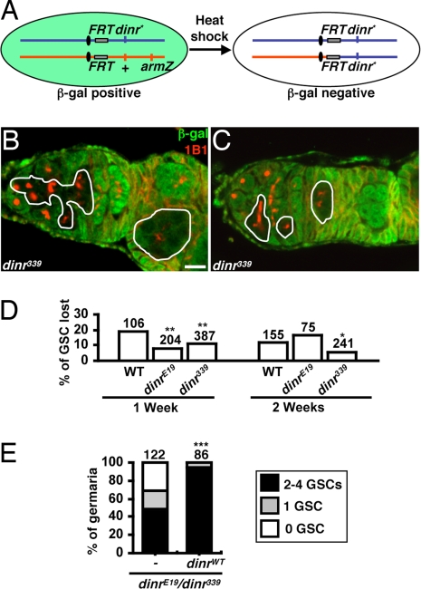
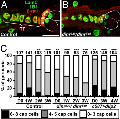
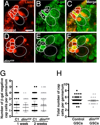
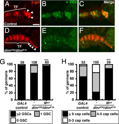
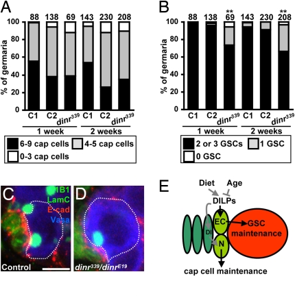
Stem cell maintenance depends on local signals provided by specialized microenvironments, or niches, in which they reside. The potential role of systemic factors in stem cell maintenance, however, has remained largely unexplored. Here, we show that insulin signaling integrates the effects of diet and age on germline stem cell (GSC) maintenance through the dual regulation of cap cell number (via Notch signaling) and cap cell-GSC interaction (via E-cadherin) and that the normal process of GSC and niche cell loss that occurs with age can be suppressed by increased levels of insulin-like peptides. These results underscore the importance of systemic factors for the regulation of stem cell niches and, thereby, of stem cell numbers.


Conflict of interest statement

The authors declare no conflict of interest.

Figures

Fig. 1.
Fig. 1.
GSC maintenance requires insulin signaling. (A) Terminal filament and cap cells form the GSC niche in Drosophila germaria. Each GSC contains a spectrosome/fusome. A GSC division generates another GSC and a cystoblast that leaves the niche and forms a 16-cell cyst enveloped by follicle cells. (B–D) One-week-old wild-type (WT) and dinr339/dinrE19 germaria labeled with vasa (red, germ cells) and 1B1 (green, fusomes). Dashed circles/ovals, GSCs. (Scale bar, 10 μm.) (E and F) GSC numbers in wild-type, dinr339/dinrE19, dinr/+ heterozygous, DILP2-overexpressing, and control females under different diets. D0, newly eclosed; Het., heterozygous; 1W, 2W, 3W, and 4W: 1-, 2-, 3-, and 4-week-old females, respectively. The number of analyzed germaria is shown above each bar. *P < 0.05, ***P < 0.001.
Fig. 2.
Fig. 2.
dinr is not required cell autonomously for GSC maintenance. (A) FLP/FRT system used to generate homozygous mutant GSCs. Flies carrying a wild-type dinr allele (+) linked to an arm-lacZ transgene in trans to a mutant or wild-type (WT) dinr allele (dinr*) were heat-shocked to induce FLP-mediated recombination between FRT sites. dinr* homozygous GSCs and their progeny are recognized by the absence of β-gal expression. (B) Mosaic germarium in which the dinr339 homozygous GSC and progeny are present. (C) Mosaic germarium in which only the dinr339 homozygous progeny are present, indicating loss of the dinr339 homozygous GSC. In B and C, solid lines outline dinr339 mutant cells. (Scale bar, 10 μm.) (D) Percentage of germaria in which the GSC has been lost. To quantify GSC loss, we calculated the percentage of germaria in which the original dinr mutant GSCs had been lost (instances equivalent to example shown in C) relative to the total number of germaria containing mosaic germline (sum of all instances equivalent to B or C). dinr mutant GSCs are not lost at a higher rate than control GSCs, showing that dinr does not promote GSC maintenance cell autonomously. The slightly lower rate of dinr339 GSC loss is consistent with findings that dinr mutant GSCs spend a higher proportion of their cell cycle displaying a round fusome morphology, which we show coincides with higher levels of E-cadherin at the cap cell–GSC junction (see text and Fig. 6 C and D). (E) GSC number in germaria of 1-week-old control dinrE19/dinr339 mutants (c587/+, CyO/+, dinrE19GAL80ts/dinr339 hh-lacZ) and those expressing wild-type dinr in germarial somatic cells (c587/+, UAS-dinrWT/+, dinrE19GAL80ts/dinr339 hh-lacZ). Somatic expression of dinr rescues the GSC loss phenotype of dinrE19/dinr339 mutants. The number of analyzed germaria is shown above each bar. *P < 0.05, **P < 0.01, **P < 0.001.
Fig. 3.
Fig. 3.
Insulin signaling controls GSC niche size. One-week-old dinr339 hh-lacZ/+ (A, control) and dinr339 hh-lacZ/dinrE19 (B) germaria labeled with LamC (green, terminal filament and cap cell nuclear envelopes), 1B1 (green, fusome), and β-gal (red, terminal filament and cap cell nuclei). Dashed ovals indicate GSCs. TF, terminal filament; *, cap cells. (Scale bar, 10 μm.) (C) Cap cell number in control, dinr339/dinrE19, and DILP2-overexpressing females (see Fig. 1 legend). The number of analyzed germaria is shown above each bar. **P < 0.01, ***P < 0.001.
Fig. 4.
Fig. 4.
Insulin receptor signaling is required in a non–cell autonomous manner to maintain cap cell numbers. Germaria from control (A–C) and dinr339 (D–F) mosaic females labeled with LamC (outlining nuclear envelope of terminal filament and cap cells) and 1B1 (outlining fusome) (A and D), β-gal (B and E), and merged (C and F). Cap cells are outlined by solid lines. GSCs are outlined by dashed ovals. Control (A–C) or dinr339 homozygous mutant (D–F) cap cell clones are recognized by the absence of β-gal (*). (Scale bar, 5 μm.) (G) Similar distribution of number of β-gal–negative cap cells per germarium in control (C1) and dinr339 mosaic females at 1 or 2 weeks after eclosion. (H) Number of cap cells in mosaic germaria containing all β-gal–negative control or dinr339 mutant GSCs in 1-week-old females, showing that dinr function is not required in GSCs to regulate the number of cap cells.
Fig. 5.
Fig. 5.
Insulin signaling maintains cap cells via Notch. Control (A–C) and dinr339 hh-lacZ/dinrE19 (D–F) germaria are labeled with β-gal (red, terminal filament and cap cell nuclei) and CD2 [green, E(spl)mβ-CD2 reporter of Notch signaling]. C and F show merge. TF, terminal filament; arrowheads, cap cells. (Scale bar, 5 μm.) GSC (G) or cap cell (H) numbers in 1-week old wild-type GAL4 controls or dinr339/dinrE19 mutants alone or expressing activated Notch (Nact). The number of germaria analyzed is shown above each bar. ***P < 0.001.
Fig. 6.
Fig. 6.
Insulin signaling within cap cells controls GSC maintenance via E-cadherin. (A) Total cap cell numbers in control mosaic germaria (C1) and in germaria without (C2) or with (dinr339) cap cell clones from dinr339 mosaics. The average cap cell number is not statistically different among genotypes. (B) Significant decrease in GSC number in germaria carrying dinr339 cap cells. The number of germaria analyzed is shown above each bar. **P < 0.01. Heterozygous (C) and dinr339/dinrE19 mutant (D) germaria labeled with 1B1 (green, fusome), LamC (green, cap cell nuclear envelopes), and E-cadherin (red). Dashed outlines indicate GSCs. (Scale bar, 5 μm.) (E) DILPs integrate age and diet effects on GSC maintenance. DILPs control niche size via Notch (N) signaling in the niche [potentially via Delta (Dl)] and cap cell–GSC association via E-cadherin (EC). Dark green, terminal filament; light green, cap cells; red, GSC.

References

    1. Li L, Xie T. Stem cell niche: Structure and function. Annu Rev Cell Dev Biol. 2005;21:605–631. - PubMed
    1. Jones DL, Wagers AJ. No place like home: Anatomy and function of the stem cell niche. Nat Rev Mol Cell Biol. 2008;9:11–21. - PubMed
    1. Pan L, et al. Stem cell aging is controlled both intrinsically and extrinsically in the Drosophila ovary. Cell Stem Cell. 2007;1:458–469. - PubMed
    1. Ryu BY, Orwig KE, Oatley JM, Avarbock MR, Brinster RL. Effects of aging and niche microenvironment on spermatogonial stem cell self-renewal. Stem Cells. 2006;24:1505–1511. - PMC - PubMed
    1. Boyle M, Wong C, Rocha M, Jones DL. Decline in self-renewal factors contributes to aging of the stem cell niche in the Drosophila testis. Cell Stem Cell. 2007;1:470–478. - PubMed

Publication types