. 2009 Jan;21(1):285-300.
doi: 10.1105/tpc.108.063248. Epub 2009 Jan 9.

The plastidial 2-C-methyl-D-erythritol 4-phosphate pathway provides the isoprenyl moiety for protein geranylgeranylation in tobacco BY-2 cells

Affiliations

The plastidial 2-C-methyl-D-erythritol 4-phosphate pathway provides the isoprenyl moiety for protein geranylgeranylation in tobacco BY-2 cells

Esther Gerber et al. Plant Cell. 2009 Jan.

Abstract

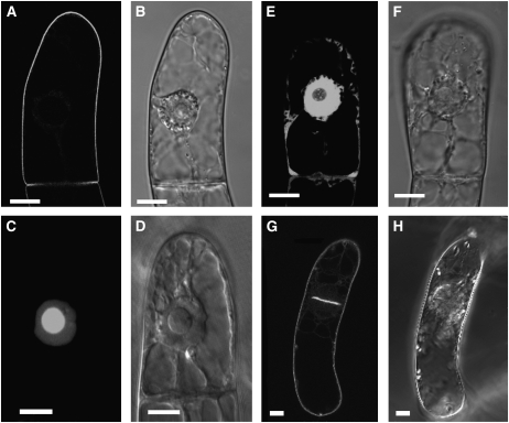
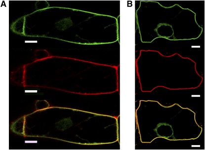
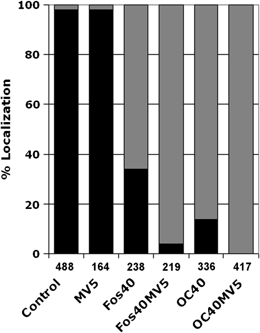
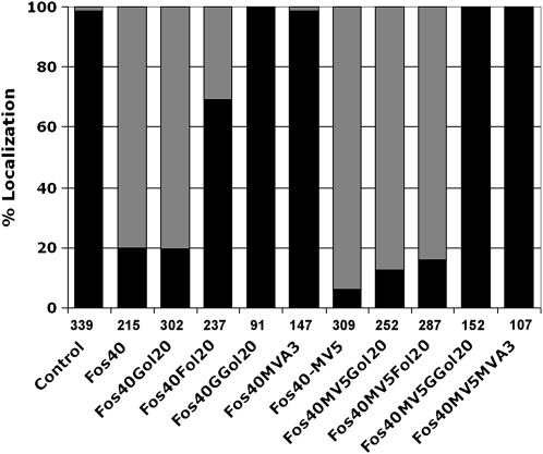
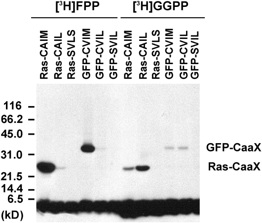
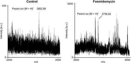
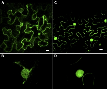
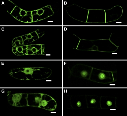
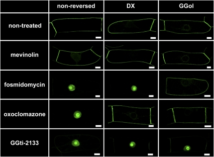
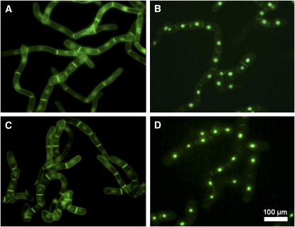
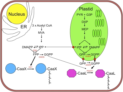
Protein farnesylation and geranylgeranylation are important posttranslational modifications in eukaryotic cells. We visualized in transformed Nicotiana tabacum Bright Yellow-2 (BY-2) cells the geranylgeranylation and plasma membrane localization of GFP-BD-CVIL, which consists of green fluorescent protein (GFP) fused to the C-terminal polybasic domain (BD) and CVIL isoprenylation motif from the Oryza sativa calmodulin, CaM61. Treatment with fosmidomycin (Fos) or oxoclomazone (OC), inhibitors of the plastidial 2-C-methyl-d-erythritol 4-phosphate (MEP) pathway, caused mislocalization of the protein to the nucleus, whereas treatment with mevinolin, an inhibitor of the cytosolic mevalonate pathway, did not. The nuclear localization of GFP-BD-CVIL in the presence of MEP pathway inhibitors was completely reversed by all-trans-geranylgeraniol (GGol). Furthermore, 1-deoxy-d-xylulose (DX) reversed the effects of OC, but not Fos, consistent with the hypothesis that OC blocks 1-deoxy-d-xylulose 5-phosphate synthesis, whereas Fos inhibits its conversion to 2-C-methyl-d-erythritol 4-phosphate. By contrast, GGol and DX did not rescue the nuclear mislocalization of GFP-BD-CVIL in the presence of a protein geranylgeranyltransferase type 1 inhibitor. Thus, the MEP pathway has an essential role in geranylgeranyl diphosphate (GGPP) biosynthesis and protein geranylgeranylation in BY-2 cells. GFP-BD-CVIL is a versatile tool for identifying pharmaceuticals and herbicides that interfere either with GGPP biosynthesis or with protein geranylgeranylation.


Figures

Figure 1.
Figure 1.
Localization of GFP Fusion Proteins to Subcellular Structures in Transformed BY-2 Cells. Stably transformed BY-2 cells in the stationary phase were diluted into fresh medium and treated under standard conditions, which are described in Results. Bars = 10 μm. (A) GFP, fused at its C terminus to the C terminus of rice calmodulin CaM61, which bears a basic domain (BD) and a CVIL geranylgeranylation motif (GFP-BD-CVIL). The resulting fluorescence is exclusively localized to the periphery of the cell. (B) The same cell as in (A) in white light mode, indicating structural details, especially the position of the nucleus. (C) Expression of a fusion protein that cannot be geranylgeranylated due to the replacement of the Cys residue in the isoprenyltransferase recognition motif by Ser (GFP-BD-SVIL). This substitution causes mislocalization of GFP-BD-CVIL to the nucleoplasm and nucleolus. (D) The same cell as in (C) in white light phase contrast mode, indicating the localization of the nucleus and the nucleolus. (E) Expression of GFP alone, with GFP fluorescence in cytoplasmic strands and nucleoplasm, but not the nucleolus. (F) The same cell as in (E) in white light mode. (G) A cell in the process of division, with accumulation of GFP-BD-CVIL in the newly forming cell plate. (H) The same cell as in (G) in white light mode.
Figure 2.
Figure 2.
Localization of GFP-BD-CVIL to the Plasma Membrane in BY-2 Cells. GFP-BD-CVIL expression was induced for 15 h with 10 μM dexamethasone before treatment with the fluorescent marker FM4-64 (6.7 μg/mL). The green channel was set for detection of GFP (top panel) and the red channel for detection of the PM marker FM4-64 (middle panel). Merged images are shown in yellow and indicate colocalization of GFP-BD-CVIL and FM4-64 (bottom panel). Bars = 10 μm. (A) In the absence of 0.23 M d-mannitol. (B) In the presence of 0.23 M d-mannitol, which induces plasmolysis. The images show single optical sections through the center of the cell, including the nucleus.
Figure 3.
Figure 3.
Quantitative Analyses of GFP-BD-CVIL Localization in BY-2 Cells Treated with MEP and MVA Pathway Inhibitors. Percentage of GFP-BD-CVIL localized to the plasma membrane and nucleus/nucleolus in the presence of MV (5 μM), Fos (40 μM), or OC (40 μM). Black bars: percentage of cells showing plasma membrane localization. Gray bars: percentage of cells with partial or complete delocalization to the nucleus/nucleolus. The number of cells analyzed is indicated under each column.
Figure 4.
Figure 4.
Chemical Complementation of Inhibitor-Induced GFP-BD-CVIL Delocalization from the Plasma Membrane to the Nucleus with MVA (3 mM) or Prenols (20 μM). Black bars: percentage of cells showing plasma membrane localization. Gray bars: percentage of cells with partial or complete delocalization to the nucleus/nucleolus. The number of cells analyzed is indicated under each column. Fos40, Fos (40 μM); MV5, MV (5 μM); Gol20, geraniol (20 μM); Fol20, farnesol (20 μM); GGol20, geranylgeraniol (20 μM); control, transformed cells without any inhibitor treatment, displaying ∼99% plasma membrane localization.
Figure 5.
Figure 5.
In Vitro Isoprenylation of Modified GFP Fusion Proteins by BY-2 Cell-Free Extracts. GFP fusion proteins were expressed in E. coli and tested for in vitro isoprenylation using cell-free extracts from 3-d-old BY-2 cells as a source of protein isoprenyltransferases and [3H]-FPP or [3H]-GGPP as isoprenyl diphosphate substrates. For comparison, Ras fusion proteins were also isoprenylated in vitro. Ras-CAIM is recognized by PFT, whereas Ras-CAIL is recognized by PGGT 1. By contrast, Ras-SVLS is not recognized by any known protein isoprenyltransferase. GFP-BD-CVIL was predicted to be a substrate of PGGT 1, whereas GFP-CVIM was predicted to be a substrate of PFT. GFP-SVIL served as a control protein that cannot be isoprenylated. The positions of GFP-CaaX and Ras-CaaX, which agree with the predicted molecular masses, are indicated. The broad band on the bottom of the gel corresponds to free radiolabeled substrate.
Figure 6.
Figure 6.
In Vivo Characterization of His6-GFP-BD-CVIL Isoprenylation by MS Analysis of His6-GFP-BD-CVIL–Derived Peptides. Solubilized total membrane and supernatant fractions from tobacco BY-2 cells induced to express His6-GFP-BD-CVIL were resolved by SDS-PAGE, and recombinant His6-GFP-BD-CVIL was cut from the gel, digested with Asp-N, and extracted and fractionated by solid-phase extraction for MALDI-TOF MS peptide mass fingerprinting and MALDI-TOF MS/MS peptide mass sequencing (detailed in Supplemental Figure 1 online). A significant peak was detected at an m/z of 2852.68, which corresponded to the predicted mass of a geranylgeranylated, methylated C-terminal His6-GFP-BD-CVIL peptide. Peaks at an m/z of 2756.62 or an m/z of 2784.56, which correspond to unprenylated and farnesylated peptides, respectively, were not detected in the absence of inhibitors (Control). However, a significant peak was detected at an m/z of 2756.62 from tobacco BY-2 cells treated with Fos.
Figure 7.
Figure 7.
Localization of GFP-CaM61 and GFP-BD-CVIL in Epidermal Cells of N. benthamiana Leaves. (A) Localization of GFP-CaM61, showing GFP fluorescence associated with the cell periphery, nucleus, and cytoplasmic strands. (B) At higher magnification, GFP-CaM61 is observed in the nucleus, but not the nucleolus. (C) Localization of GFP-BD-CVIL, showing GFP fluorescence associated with the plasma membrane and nucleus, but not cytoplasmic strands. (D) At higher magnification (threefold electronic zoom), GFP-BD-CVIL is observed to be concentrated in the nucleolus. Bars = 10 μm. [See online article for color version of this figure.]
Figure 8.
Figure 8.
Localization of GFP-CaM61 and GFP-BD-CVIL in BY-2 Cells. (A) Localization of GFP-CaM61 in BY-2 cells, showing GFP fluorescence associated with ER and perinuclear membranes, plasma membrane, and cytoplasmic strands. (B) Localization of GFP-BD-CVIL in BY-2 cells, showing GFP fluorescence associated exclusively with the plasma membrane. (C) Localization of GFP-CaM61 in BY-2 cells treated with MV as described by Dong et al. (2002), showing GFP fluorescence associated with ER and perinuclear membranes, plasma membrane, and cytoplasmic strands. (D) Localization of GFP-BD-CVIL in BY-2 cells treated with MV as described by Dong et al. (2002), showing GFP fluorescence associated with the plasma membrane (faint fluorescence is also seen in the nucleus). (E) Localization of GFP-CaM61 in BY-2 cells treated with Fos, showing partial translocation of GFP fluorescence to the nucleus, but not the nucleolus. (F) Localization of GFP-BD-CVIL in BY-2 cells treated with Fos, showing translocation of GFP fluorescence to the nucleus and nucleolus. (G) Localization of GFP-CaM61 in BY-2 cells treated with MV and Fos, showing increased translocation of GFP fluorescence to the nucleus. (H) Localization of GFP-BD-CVIL in BY-2 cells treated with MV and Fos, showing almost complete translocation of GFP fluorescence to the nucleus and nucleolus. Bars = 10 μm. [See online article for color version of this figure.]
Figure 9.
Figure 9.
A Bioassay for the Identification of MEP Pathway and PGGT 1 Inhibitors. His6-tagged GFP-BD-CVIL localization in transformed BY-2 cells. His6-GFP-BD-CVIL was localized to the periphery of transformed BY-2 cells in the absence of inhibitors. MV (5 μM) exerted no detectable effect on His6-GFP-BD-CVIL localization. By contrast, Fos (40 μM) caused a dramatic translocation of His6-GFP-BD-CVIL to the nucleus/nucleolus. GGol (5 μM), but not DX (0.5 mM), reversed this effect (Fos inhibits MEP synthase). OC (10 μM) also caused a dramatic translocation of His6-GFP-BD-CVIL to the nucleus/nucleolus, but GGol and DX reversed this effect (OC inhibits DXS). In addition, GGti-2133 (30 μM) caused His6-GFP-BD-CVIL to accumulate in the nucleus/nucleolus, but was not reversed by GGol or DX (GGti-2133 inhibits PGGT 1). Bars = 10 μm. [See online article for color version of this figure.]
Figure 10.
Figure 10.
Reproducibility of Effects on a Random Collection of BY-2 Cells. Fluorescence images at low resolution of large numbers of cloned BY-2 cells expressing His6-GFP-BD-CVIL (Nikon DXM11200 CCD color camera, 20 × 0.45 objective; filters: EX460-500, DM505, EM510-560). [See online article for color version of this figure.] (A) Untreated cells. (B) to (D) Cells treated with 30 μM OC (B), 5 μM MV (C), and 30 μM OC plus 5 μM MV (D).
Figure 11.
Figure 11.
Biosynthetic Origins of Farnesyl Diphosphate and Geranylgeranyl Diphosphate in BY-2 Cells for Protein Farnesylation and Geranylgeranylation, Respectively. CaaX, isoprenylation motif; DMAPP, dimethyl allyl diphosphate; DXP, 1-deoxy-d-xylulose 5-phosphate; FDS, farnesyl diphosphate synthase; GGDS, geranylgeranyl diphosphate synthase; G3P, d-glyceraldehyde 3-phosphate; GPP, geranyl diphosphate; IPP, isopentenyl diphosphate; PYR, pyruvate.

Comment in

  • A role for plastids in plant protein isoprenylation.
    Crowell DN, Hemmerlin A, Gerber E, Hartmann M, Heintz D, Rohmer M, Bach TJ. Crowell DN, et al. Plant Signal Behav. 2009 Mar;4(3):217-8. doi: 10.4161/psb.4.3.7842. Plant Signal Behav. 2009. PMID: 19721754 Free PMC article.

References

    1. Adam, K.-P., Thiel, R., and Zapp, J. (1999). Incorporation of 1-[1-13C]deoxy-D-xylulose in chamomile sequiterpenes. Arch. Biochem. Biophys. 369 127–132. - PubMed
    1. Alberts, A.W., et al. (1980). Mevinolin, a highly potent competitive inhibitor of hydroxymethylglutaryl-coenzyme A reductase and a cholesterol-lowering agent. Proc. Natl. Acad. Sci. USA 77 3957–3961. - PMC - PubMed
    1. Aoyama, T., and Chua, N.H. (1997). A glucocorticoid-mediated transcriptional induction system in transgenic plants. Plant J. 11 605–612. - PubMed
    1. Bach, T.J. (1995). Some new aspects of isoprenoid biosynthesis in plants - A review. Lipids 30 191–202. - PubMed
    1. Bach, T.J., and Lichtenthaler, H.K. (1983). Inhibition by mevinolin of plant growth, sterol formation and pigment accumulation. Physiol. Plant. 59 50–60.

Publication types

MeSH terms

Associated data

LinkOut - more resources