. 2009 May;50(5):832-45.
doi: 10.1194/jlr.M800402-JLR200. Epub 2009 Jan 9.

Oxidized LDL impair adipocyte response to insulin by activating serine/threonine kinases

Affiliations

Oxidized LDL impair adipocyte response to insulin by activating serine/threonine kinases

Beatrice Scazzocchio et al. J Lipid Res. 2009 May.

Abstract

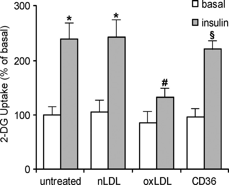
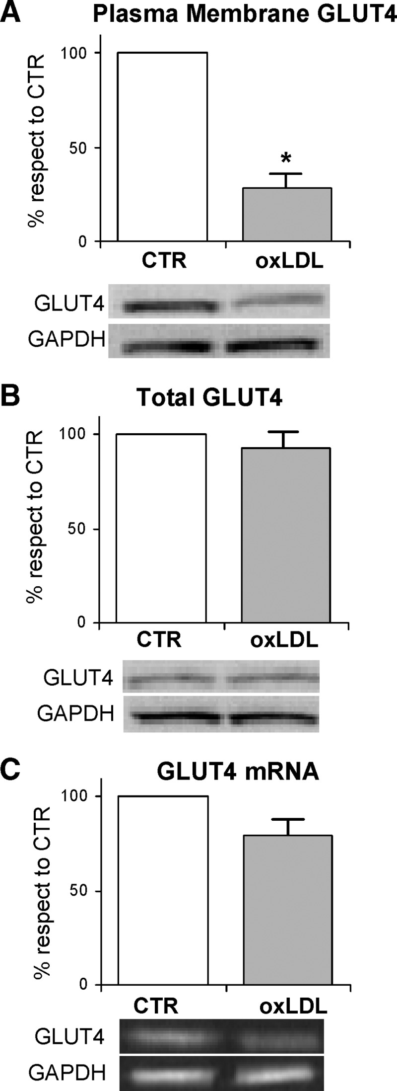
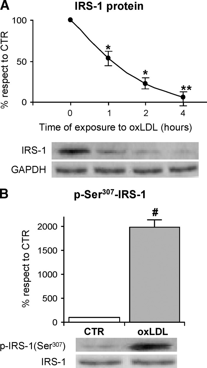
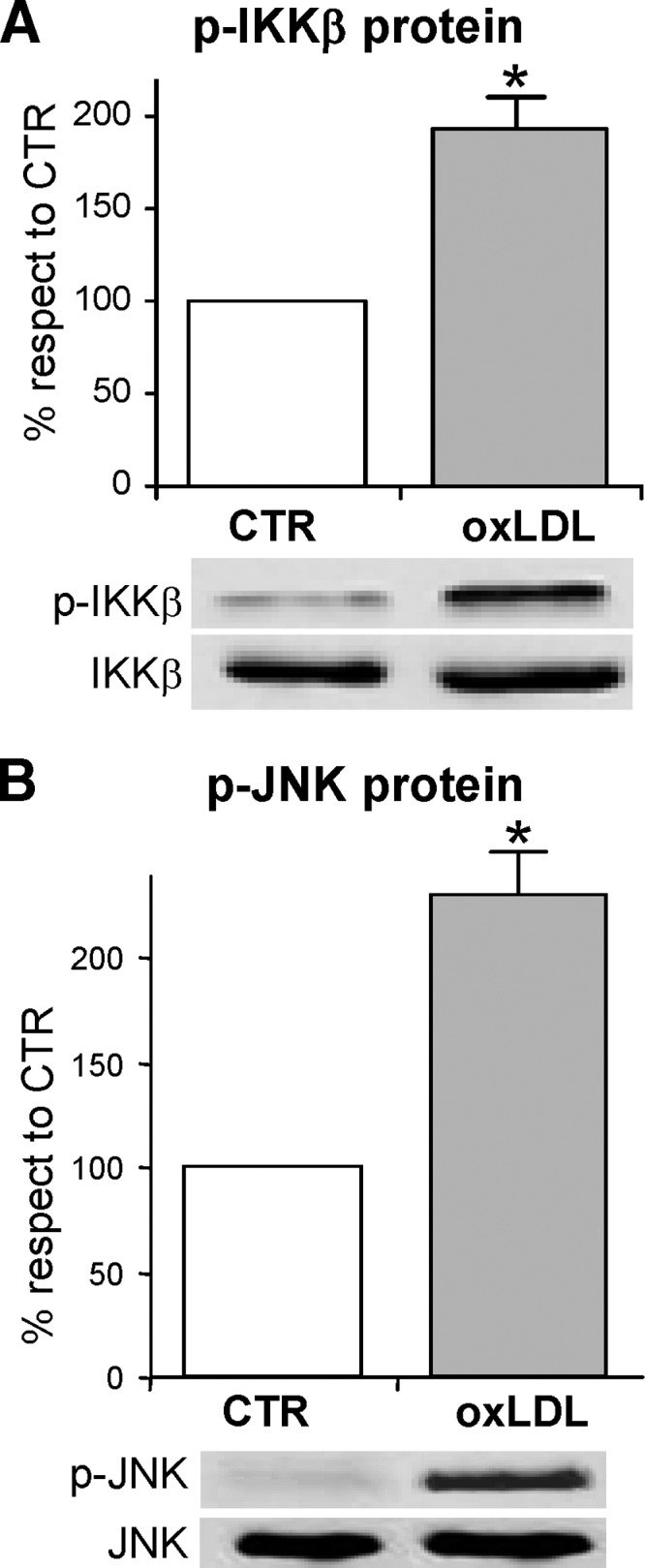
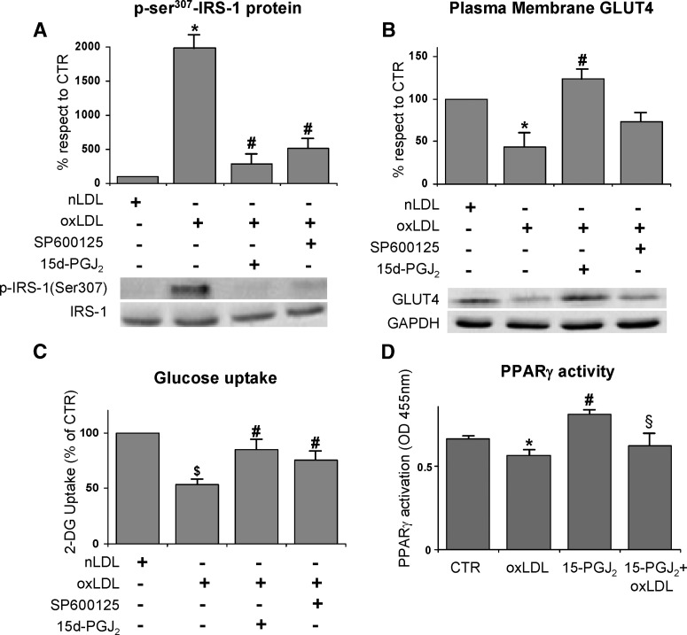
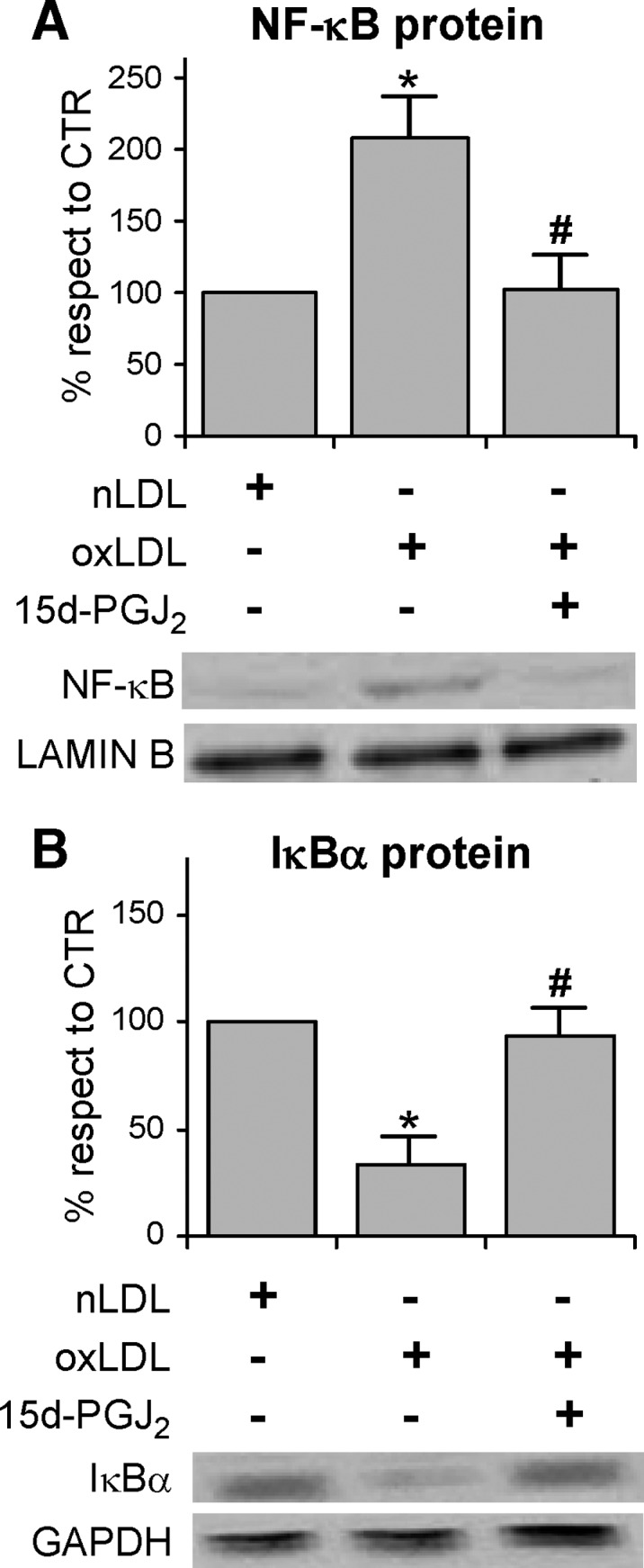
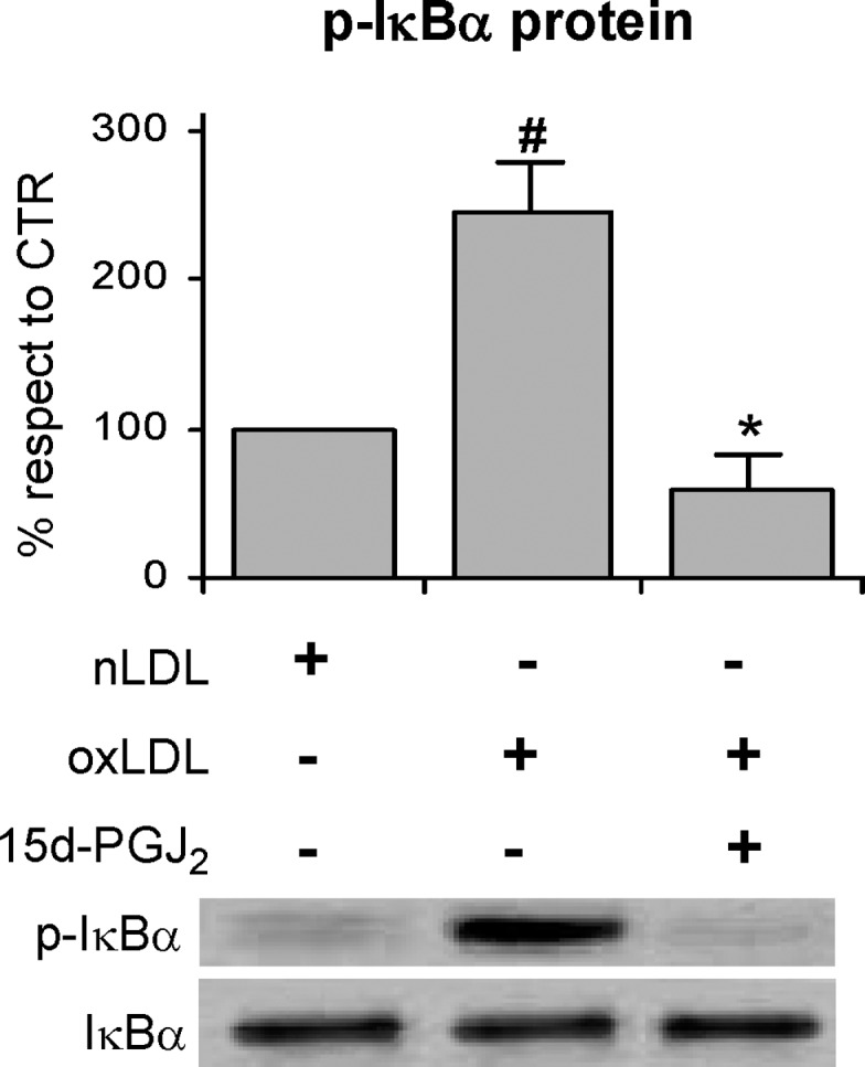
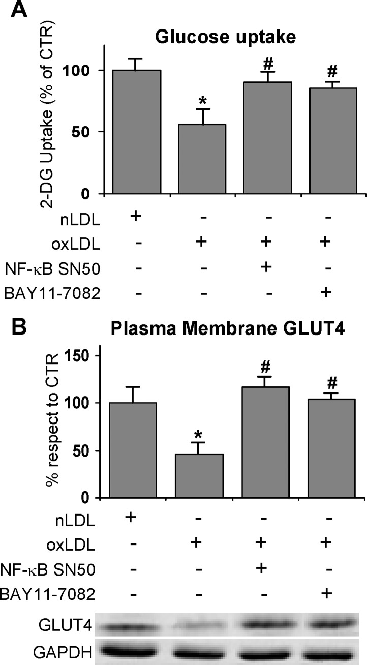
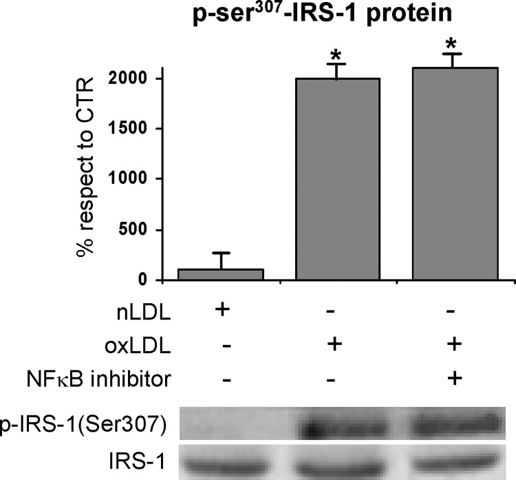
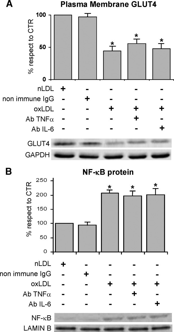
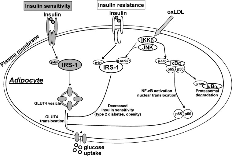
Oxidized LDL (oxLDL) increase in patients affected by type-2 diabetes, obesity, and metabolic syndrome. Likewise, insulin resistance, an impaired responsiveness of target tissues to insulin, is associated with those pathological conditions. To investigate a possible causal relationship between oxLDL and the onset of insulin resistance, we evaluated the response to insulin of 3T3-L1 adipocytes treated with oxLDL. We observed that oxLDL inhibited glucose uptake (-40%) through reduced glucose transporter 4 (GLUT4) recruitment to the plasma membrane (-70%), without affecting GLUT4 gene expression. These findings were associated to the impairment of insulin signaling. Specifically, in oxLDL-treated cells insulin receptor (IR) substrate-1 (IRS-1) was highly degraded likely because of the enhanced Ser(307)phosphorylation. This process was largely mediated by the activation of the inhibitor of kappaB-kinase beta (IKKbeta) and the c-Jun NH(2)-terminal kinase (JNK). Moreover, the activation of IKKbeta positively regulated the nuclear content of nuclear factor kappaB (NF-kappaB), by inactivating the inhibitor of NF-kappaB (IkappaBalpha). The activated NF-kappaB further impaired per se GLUT4 functionality. Specific inhibitors of IKKbeta, JNK, and NF-kappaB restored insulin sensitivity in adipocytes treated with oxLDL. These data provide the first evidence that oxLDL, by activating serine/threonine kinases, impaired adipocyte response to insulin affecting pathways involved in the recruitment of GLUT4 to plasma membranes (PM). This suggests that oxLDL might participate in the development of insulin resistance.


Figures

Fig. 1.
Fig. 1.
Oxidized LDL (OxLDL) decrease glucose uptake in 3T3-L1 adipocytes after internalization by CD36 3T3-L1 adipocytes were serum starved in low-glucose medium for 18 h, preincubated with 10 μg/ml monoclonal anti-CD36 antibody for 30 min, treated with 100 mg/l native LDL (nLDL) or oxLDL for 4 h before incubation with 20 nM insulin for 15 min. The rate of glucose uptake was determined upon addition of [H3]2-DG (2-deoxyglucose) for 45 min. Basal glucose uptake in untreated cells was set at 100% to which all other values were related. The data are means ± SEM of three independent experiments. * P < 0.05 compared with the basal uptake; # P < 0.05 compared with untreated or nLDL treated cells; § P < 0.05 compared with oxLDL treated cells.
Fig. 2.
Fig. 2.
Gene and protein expressions of glucose transporter 4 (GLUT4) in oxLDL-treated adipocytes 3T3-L1 adipocytes were serum starved for 18 h and incubated with 100 mg/l nLDL or oxLDL for 4 h before incubation with 20 nM insulin for 15 min. A: Plasma membrane fractions were prepared as described in Materials and Methods. Immunoblotting of GLUT4 and GAPDH were determined. B: The cell lysates were resolved by SDS-PAGE and analyzed using antibodies against GLUT4 and GAPDH. The blots are representative of at least three independent experiments. C: Total RNA was analyzed by RT-PCR as described in Materials and Methods. The values indicate expression of target gene normalized to GAPDH RNA and expressed as percentage of nLDL control (CTR). The data are means of three independent experiments ± SEM. * P < 0.001 compared with nLDL (CTR).
Fig. 3.
Fig. 3.
Immunoblotting and serine phosphorylation of insulin receptor substrate (IRS)-1 3T3-L1 adipocytes were serum-starved for 18 h and treated with 100 mg/l nLDL or oxLDL from 30 min to 4 h and then stimulated with 20 nM insulin for 15 min. A: Total cell lysates were separated by SDS-PAGE and analyzed using anti-IRS-1 antibody. Results were normalized to GAPDH protein content. B: Immunoprecipitates with anti-IRS-1 antibody were separated by 7.5% SDS-PAGE, and analyzed with antinonphospho- or antiphospho-ser307 IRS-1 antibody as described in Materials and Methods. Blots are representative of at least three independent experiments at 1 h. The data are means ± SEM of three independent experiments. * P < 0.001 compared with time 0; # P < 0.001 compared with nLDL (CTR).
Fig. 4.
Fig. 4.
Activation of the Serine Kinases by oxLDL Activations of inhibitor of κB-kinase β (IKKβ) (A) and c-Jun NH2-terminal kinase (JNK) (B) by oxLDL are shown. 3T3-L1 adipocytes were serum-starved for 18 h, treated with 100 mg/l nLDL or oxLDL for 1 h and stimulated with 20 nM insulin for 15 min. Immunoprecipitates with anti-IKKα/β or anti-SAPK/JNK were separated by 12% SDS-PAGE and analyzed using antinonphospho- or phosphoantibodies against IKKα/β (A) or JNK (B), as described in Materials and Methods. Representative images are shown from three independent experiments. * P < 0.05 compared with nLDL (CTR).
Fig. 5.
Fig. 5.
Effects of specific kinase inhibitors on oxLDL-stimulated insulin resistance 3T3-L1 adipocytes were serum starved for 18 h, preincubated with 50 μM 15-deoxy-Δ12,14-prostaglandin J2 (15d-PGJ2) or 50 μM SP600125 for 30 min, treated with 100 mg/l nLDL or oxLDL, and then stimulated with 20 nM insulin for 15 min. A: Effects of IKKβ and JNK inhibitors on oxLDL-induced serine phosphorylation of IRS-1. The immunoprecipitates with IRS-1 antibody were resolved by 7.5% SDS-PAGE and analyzed with antinonphospho- or antiphospho-ser307 IRS-1 antibody. Blots are representative of at least three independent experiments at 1 h. B: Immunoblotting of GLUT4 on plasma membrane fractions. Cells were washed after nLDL or oxLDL treatment for 4 h, and plasma membrane fractions were prepared as described in Materials and Methods. Representative blots from at least four independent experiments are shown. Results were normalized to GAPDH protein content. C: Effects of IKKβ and JNK inhibitors on oxLDL-inhibited glucose uptake. The rate of glucose uptake was determined upon addition of [H3]2-DG for 45 min. D: Effect of IKKβ inhibitor on PPARγ activation. The data are means ± SEM of three independent experiments. * P < 0.001 compared with nLDL (CTR); # P < 0.05 compared with oxLDL; $ P < 0.05 compared with nLDL (CTR); § P < 0.05 compared with 15-PGJ2 treated cells.
Fig. 6.
Fig. 6.
Effects of IKKβ inhibitor on nuclear factor κB (NF-κB) activation and IκBα degradation in oxLDL-induced 3T3-L1 adipocytes Cells were serum starved for 18 h, preincubated with 50 μM 15d-PGJ2 for 30 min, treated with 100 mg/l nLDL or oxLDL for 4 h, and then stimulated with 20 nM insulin for 15 min. Nuclear and cytosolic extracts were prepared as described in Materials and Methods and analyzed by immunoblotting of NF-κB p65 (A), IκBα (B). Lamin B and GAPDH antibodies were used as markers for nuclear and cytosolic extracts, respectively. Representative images are shown from three independent experiments. * P < 0.05 compared with nLDL (CTR). # P < 0.05 compared with oxLDL.
Fig. 7.
Fig. 7.
IKKβ inhibitor prevents oxLDL-induced IκBα degradation in 3T3-L1 adipocytes 3T3-L1 adipocytes were preincubated with 50 μM 15d-PGJ2 for 30 min followed by nLDL or oxLDL treatment for 4 h before stimulation with 20 nM insulin for 15 min as described in Materials and Methods. Total cell lysates were analyzed by Western blotting using anti-phospho(ser32/36)-IκBα and anti-GAPDH antibodies. Representative images are shown from three independent experiments. # P < 0.05 compared with nLDL (CTR); * P < 0.001 compared with oxLDL.
Fig. 8.
Fig. 8.
Effects of NFκB inhibitors on oxLDL treatment 3T3-L1 adipocytes were serum starved for 18 h, preincubated with 10 μM SN50 or 5μM BAY-11-7082 for 30 min, treated with 100 mg/l nLDL or oxLDL, and then stimulated with 20 nM insulin for 15 min. A: Effects of NF-κB inhibitors on oxLDL-inhibited glucose uptake. Cells were washed after 4 h of nLDL or oxLDL treatment. The rate of glucose uptake was determined upon addition of [H3]2-DG for 45 min. B: Immunoblotting of GLUT4 on plasma membrane fractions. Cells were washed after nLDL or oxLDL treatment for 4 h and plasma membrane fractions were prepared as described in Materials and Methods. Results were normalized to GAPDH protein content. Blots are representative of at least four independent experiments. The data are means ± SEM of three independent experiments. * P < 0.05 compared with nLDL (CTR); # P < 0.05 compared with oxLDL.
Fig. 9.
Fig. 9.
Effects of NFκB inhibitor on oxLDL-induced serine phosphorylation of IRS-1. Cells were prepared after 1 h treatment with nLDL or oxLDL, and immunoprecipitates with IRS-1 antibody were resolved by 7.5% SDS-PAGE and analyzed with antinonphospho- or antiphospho-ser307 IRS-1 antibody. Blots are representative of at least four independent experiments. The data are means of four independent experiments ± SEM.* P < 0.001 compared with nLDL (CTR).
Fig. 10.
Fig. 10.
Effects of anti-TNFα and anti-IL-6 neutralizing antibodies on GLUT4 membrane translocation and NF-κB activation in oxLDL-treated 3T3-L1 adipocytes. 3T3-L1 adipocytes were serum starved for 18 h, preincubated with monoclonal anti-TNFα (2 μg/ml) or anti-IL-6 (0.5 μg/ml) antibodies for 30 min, treated with 100 mg/l nLDL or oxLDL, and then stimulated with 20 nM insulin for 15 min. A: Immunoblotting of GLUT4 on plasma membrane fractions. Plasma membrane fractions were prepared as described in Materials and Methods. Results were normalized to GAPDH protein content. B: Immunoblotting of NF-κB p65 on nuclear extracts. Nuclear extracts were prepared as described in Materials and Methods and analyzed by immunoblotting of NF-κB p65. Lamin B antibody was used as marker for nuclear extracts.
Fig. 11.
Fig. 11.
Schematic representation of the oxLDL effects on insulin sensitivity in 3T3-L1 adipocytes. OxLDL increase serine307-phosphorylation of IRS-1 by IKKβ and JNK. The impairment of insulin signaling leads to reduced uptake of glucose via GLUT4. IKKβ is also involved in the activation of NF-κB signaling pathway by phosphorylating IκBα. NF-κB downregulates GLUT4 translocation and glucose uptake. OxLDL, oxidized lipoproteins; IRS-1, insulin receptor substrate 1; GLUT4, glucose transporter 4; p-tyr, phosphotyrosine; p-ser, phosphoserine; IKKβ, inhibitory protein κB kinase β; JNK, c-Jun NH2-terminal kinase; IκBα, inhibitory κB protein α; NF-κB, nuclear factor κB.

References

    1. Pradhan A. 2007. Obesity, metabolic syndrome, and type 2 diabetes: inflammatory basis of glucose metabolic disorders. Nutr. Rev. 65 S152–S156. - PubMed
    1. Guilherme A., J. V. Virbasius, V. Puri, and M. P. Czech. 2008. Adipocyte dysfunctions linking obesity to insulin resistance and type 2 diabetes. Nat. Rev. Mol. Cell Biol. 9 367–377. - PMC - PubMed
    1. Hegele R. A. 2000. Familial partial lipodystrophy: a monogenic form of the insulin resistance syndrome. Mol. Genet. Metab. 71 539–544. - PubMed
    1. Kim J. K., O. Gavrilova, Y. Chen, M. L. Reitman, and G. I. Shulman. 2000. Mechanism of insulin resistance in A-ZIP/F-1 fatless mice. J. Biol. Chem. 275 8456–8460. - PubMed
    1. Gan S. K., K. Samaras, A. Carr, and D. Chisholm. 2001. Anti-retroviral therapy, insulin resistance and lipodystrophy. Diabetes Obes. Metab. 3 67–71. - PubMed

MeSH terms