. 2009 Mar 27;284(13):8427-38.
doi: 10.1074/jbc.M805582200. Epub 2009 Jan 9.

Type XIV Collagen Regulates Fibrillogenesis: PREMATURE COLLAGEN FIBRIL GROWTH AND TISSUE DYSFUNCTION IN NULL MICE

Affiliations

Type XIV Collagen Regulates Fibrillogenesis: PREMATURE COLLAGEN FIBRIL GROWTH AND TISSUE DYSFUNCTION IN NULL MICE

Heather L Ansorge et al. J Biol Chem. .

Abstract

Type XIV collagen is a fibril-associated collagen with an interrupted triple helix. This collagen interacts with the fibril surface and has been implicated as a regulator of fibrillogenesis; however, a specific role has not been elucidated. Functional roles for type XIV collagen were defined utilizing a new type XIV collagen-deficient mouse line. This line was produced using a conventional targeted knock-out approach. Col14a1(-/-) mice were devoid of type XIV collagen, whereas heterozygous mice had reduced synthesis. Both mutant Col14a1 genotypes were viable with a grossly normal phenotype; however, mature skin exhibited altered mechanical properties. Prior to evaluating tendon fibrillogenesis in type XIV collagen-deficient mice, the developmental expression patterns were analyzed in wild-type flexor digitorum longus (FDL) tendons. Analyses of mRNA and protein expression indicated tissue-specific temporal expression that was associated with the early stages in fibrillogenesis. Ultrastructural analyses of wild-type and null tendons demonstrated premature fibril growth and larger fibril diameters in tendons from null mice at postnatal day 4 (P4). However, fibril structure in mature tendons was normal. Biomechanical studies established a direct structure/function relationship with reduced strength in P7-null tendons. However, the biomechanical properties in P60 tendons were comparable in null and wild-type mice. Our results indicate a regulatory function for type XIV collagen in early stages of collagen fibrillogenesis with tissue differences.


Figures
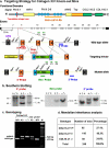
FIGURE 1.
FIGURE 1.
Creation of Col14a1 knock-out mice. a, targeting strategy. Functional domains of type XIV collagen and genomic structure of mouse Col14a1 are shown in the upper part of the diagram. In the gene-targeting vector, exon 4 was split into two parts, and a stop codon was inserted into the 5′-half of E4 by PCR. Genomic sequences of exon 3 through the first part of exon 4 and the second part of exon 4 through exon 5 were chosen as 5′- and 3′-targeting arms. Both arms are about 3 kb in size. Neomycin-resistant gene was inserted between the two arms, reversely (black arrows show transcription direction). Probes (short color rectangles) and restriction enzymes used for genomic DNA digestion and Southern blotting are indicated in the diagram. Green arrows represent gene-specific primers for genotyping. Primer 1 (P1) and Primer 2 (P2), from the second part of exon 4 and intron 4, respectively, produce a PCR product from the wild-type and mutant alleles. Primer 3 (P3 from the Neo gene, produces a DNA fragment only from the mutant allele when used with P2. The translation termination codon, which is located in FNIII-1, is marked on the protein structure diagram. The anti-type XIV collagen antibody epitope also is marked in this diagram. b, genotype analysis of Col14a1 knock-out mice using Southern blotting. A 9.9-kb and a 7.2-kb DNA fragment corresponding to the wild-type and null alleles, respectively, were detected by 5′-probe hybridized with XbaI digested mouse genomic DNA. Neo probe was used for hybridization with SpeI-digested DNA and a 10.4-kb DNA fragment was detected. A 6.3-kb DNA fragment indicating the wild-type allele and a 7.5-kb DNA fragment indicating the mutant allele were recognized by 3′-probe after hybridization with EcoRV-digested DNA. c, PCR genotyping. A 410-bp wild-type allele PCR product and a 540-bp mutant allele PCR product were generated by PCR genotyping, which identified the three Col14a1 genotypes. Col14a1(+/–) ES cell and wild-type C57 mouse genomic DNA samples were used as controls. d, Mendelian inheritance analysis. A total of 240 offspring from heterozygous Col14a1 breeders were genotyped. The ratio of wild type, heterozygous, and homozygous null mice is roughly 1:2:1.
FIGURE 2.
FIGURE 2.
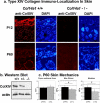
Absence of type XIV collagen and alteration of biomechanical properties in Col14a1-null mouse skin. a, immunofluorescence staining of Col14a1 knock-out mouse skin using anti-type XIV collagen antibody was completely negative in Col14a1-null mice skin. DAPI staining shows skin structure (blue nuclear). b, Western blotting of protein samples extracted from mouse skin showed complete absence of type XIV collagen in Col14a1-null mice and reduced synthesis in heterozygous mice. c, biomechanical studies showed a significant decrease in the maximum stress in the P60 skin of the col14a1(–/–) mice compared with that of wild-type controls. Additionally, the modulus showed a trend toward lower modulus when compared with wild-type mice. No significant differences were found in the cross-sectional area of the P60 skin. *, p < 0.05; #, p < 0.1.
FIGURE 3.
FIGURE 3.
Type XIV collagen expression in normal developing mouse tendons. Type XIV collagen mRNA expression (between P4-P90) was analyzed by real time quantitative RT-PCR (a) and semi-quantitative RT-PCR (b). A mixture of 2 cDNA preparations from samples at each stage was used as a template for PCR. The PCR was done in triplicate, and the results are expressed relative to 18S mRNA amplified in parallel. c, semi-quantitative immunoblotting analysis. Actin reactivity was used to monitor comparable protein loading. All results demonstrate a clear decrease in type XIV collagen in development.
FIGURE 4.
FIGURE 4.
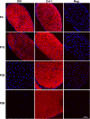
Type XIV collagen expression patterns in mouse tendon development. Localization of type XIV collagen or type I collagen by immunofluorescence microscopy with nuclear localization using DAPI (blue) is shown. There was a dramatic decrease in reactivity against type XIV collagen between P10 and P30 whereas reactivity against type I collagen was comparable at P4 through P90. The spatial localization of both types XIV and I collagen were comparable at P4 and P10 consistent with interactions during fibrillogenesis. Type XIV collagen was present at early stages (P4 and 10) and virtually absent at later stages (P30 and 90) stages in tendon development, consistent with the mRNA and protein data. Negative controls were secondary antibody alone and images captured at settings identical to those for type XIV collagen.
FIGURE 5.
FIGURE 5.
Altered fibroblast and fiber organization in P4 tendons from Col14a1(–/–) mice. Transmission electron micrographs of FDL tendons at postnatal day 4 (P4) and P60 from Col14a1(+/+) and Col14a1(–/–) mice. At P4, tendon fibroblasts are surrounded by small collagen fibers (fibril bundles) (*). The wild-type collagen fibers are uniform in shape, whereas the mutant bundles are more irregular. In addition, the fibroblast and the compartments defined by their processes are disrupted in the mutant tendon relative to the organized wild-type controls. At P60, the collagen fibers are much larger than at P4 are defined by attenuated fibroblast processes (arrows). There is comparable structure and organization in Col14a1(+/+) and Col14a1(–/–) tendons at this mature time point when type XIV collagen is not expressed in wild-type mice. Bar, 3 μm.
FIGURE 6.
FIGURE 6.
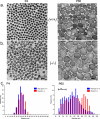
Altered collagen fibril formation in Col14a1(–/–) tendons. Transmission electron micrographs of FDL tendon at postnatal day 4 (P4) from (a) Col14a1(+/+) and (b) Col14a1(–/–) mice. At P4, the fibrils of the wild-type control are more uniform in shape and smaller than the fibrils in the Col14a1(–/–) tendon, which are more irregular and larger. At P60, the mature fibrils from the wild-type tendon are of variable size with small to larger cross sectional profiles. In the Col14a1(–/–) tendon, there are fewer smaller fibrils and more fibrils of larger diameter as compared with the wild-type control. Bar, 100 nm. c, histograms representing fibril diameter measurements of tendons at P4 and P60. In the P4 wild-type tendon, the fibril diameter distribution demonstrates a unimodal pattern with the majority of fibril diameters between 35 and 45 nm. In the P4 mutant tendon, the fibril diameter measurements also show a unimodal (50–60 nm) distribution; however, there is a greater frequency of larger diameter fibrils. At P60, the wild-type diameter measurements have a much broader multimodal distribution. The fibril diameter measurements in the mutant tendon also have a multimodal distribution. In contrast to the wild type distribution, there is a reduced frequency of smaller diameter fibrils in the null tendons. In addition, a distinct subpopulation of larger diameter fibrils, absent in the wild-type tendons, is present in the null tendons. We suggest that this represents the fibrils that prematurely enter the fibril growth phase in the absence of type XIV collagen.
FIGURE 7.
FIGURE 7.
Tendon biomechanics in the Col14a1-null mouse. a, P7 FDLs. Col14a1(–/–) mice demonstrated a significant reduction in maximum load, stiffness, and modulus when compared with wild-type tendons. In addition, FDLs from the developing Col14a1(–/–) mice showed significantly decreased maximum load and a trend toward decreased maximum stress and stiffness when compared with the heterozygote mice. FDLs from Col14a1(+/–) mice also had significantly decreased stiffness when compared with wild-type mice. b, P60 FDLs. The mature tendons demonstrated no significant differences between the Col14a1(+/+), Col14a1(+/–), and Col14a1(–/–) tendons. *, p < 0.05; #, p < 0.1.
FIGURE 8.
FIGURE 8.
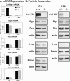
Leucine-rich proteoglycan expression in the Col14a1-null mouse. a, mRNA expression in P4 and P30 FDLs. Type XIV collagen, biglycan, decorin, lumican, and fibromodulin mRNA expression were analyzed by real time RT-PCR. Actin was used as an endogenous control to standardize the amount of sample RNA. The results shown represent three independent experiments. Error bars represent the pooled standard deviation. Col14a1(–/–) mice demonstrated no significant changes in mRNA expression between wild-type and Col14a1(–/–) mice (p > 0.1). This was the case for biglycan, decorin, lumican, and fibromodulin at both developmental stages. b, semi-quantitative immunoblot analysis. Extract (10 mg) prepared from P4 and P30 FDLs was subjected to immunoblot analysis with antibodies specified to collagen type XIV, biglycan, decorin, lumican, fibromodulin, and actin. Actin reactivity was used as loading control. Relative expression levels of indicated proteoglycan showed that the core protein expression in the wild type compared with null mice was comparable for all four proteoglycans at both developmental stages.

References

    1. van der Rest, M., and Garrone, R. (1991) FASEB J. 5 2814–2823 - PubMed
    1. Kielty, C. M., and Grant, M. E. (2002) in Connective Tissue and Its Heritable Disorders (Royce, P. M., and Steinmann, B., eds) pp. 159–222, Wiley-Liss, New York
    1. Birk, D. E., and Bruckner, P. (2005) in Topics in Current Chemistry: Collagen (Brinckmann, J., Müller, P. K., and Notbohm, H., eds) pp. 185–205, Springer-Verlag, Berlin
    1. Birk, D. E., Nurminskaya, M. V., and Zycband, E. I. (1995) Dev. Dyn. 202 229–243 - PubMed
    1. Birk, D. E., and Mayne, R. (1997) Eur. J. Cell Biol. 72 352–361 - PubMed

Publication types

LinkOut - more resources