. 2009 Jan 29;457(7229):557-61.
doi: 10.1038/nature07665. Epub 2009 Jan 11.

Adaptive immune features of natural killer cells

Affiliations

Adaptive immune features of natural killer cells

Joseph C Sun et al. Nature. .

Erratum in

  • Nature. 2009 Feb 26;457(7233):1168

Abstract

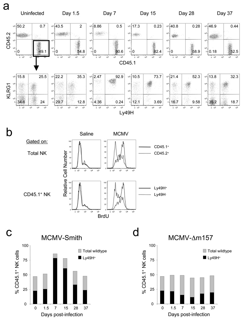
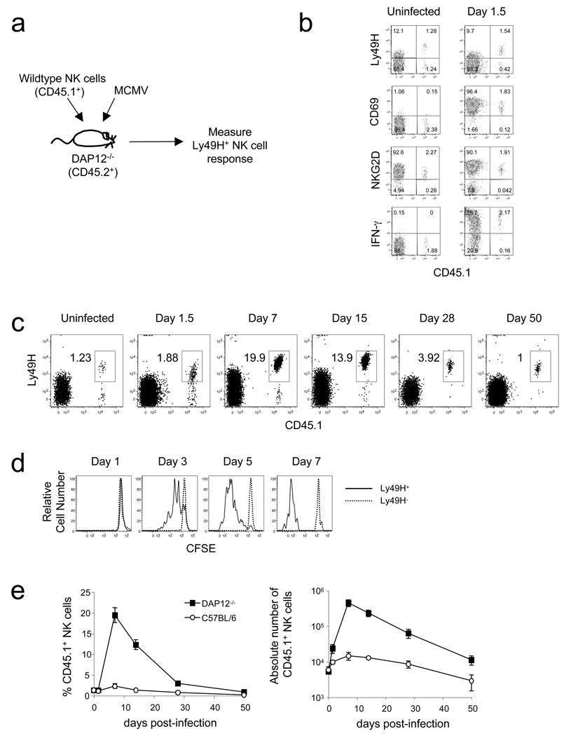
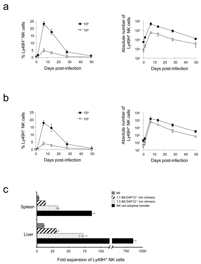
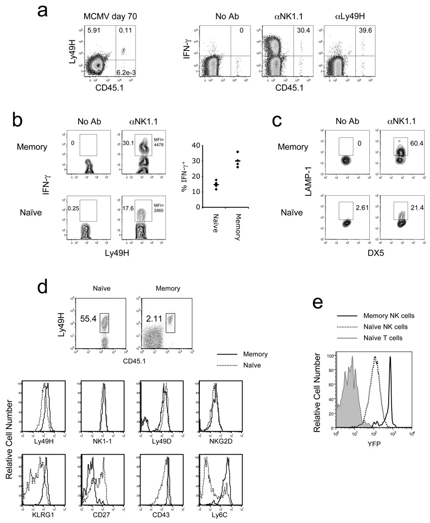
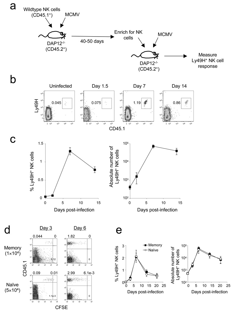
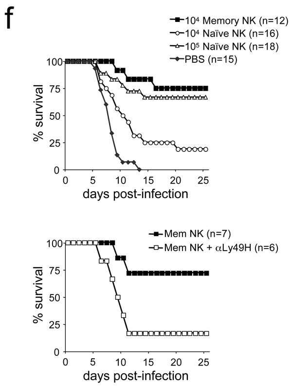
In an adaptive immune response, naive T cells proliferate during infection and generate long-lived memory cells that undergo secondary expansion after a repeat encounter with the same pathogen. Although natural killer (NK) cells have traditionally been classified as cells of the innate immune system, they share many similarities with cytotoxic T lymphocytes. We use a mouse model of cytomegalovirus infection to show that, like T cells, NK cells bearing the virus-specific Ly49H receptor proliferate 100-fold in the spleen and 1,000-fold in the liver after infection. After a contraction phase, Ly49H-positive NK cells reside in lymphoid and non-lymphoid organs for several months. These self-renewing 'memory' NK cells rapidly degranulate and produce cytokines on reactivation. Adoptive transfer of these NK cells into naive animals followed by viral challenge results in a robust secondary expansion and protective immunity. These findings reveal properties of NK cells that were previously attributed only to cells of the adaptive immune system.


Figures

Figure 1
Figure 1. Preferential expansion of wild-type but not DAP12-deficient NK cells during MCMV infection
Mixed bone marrow chimeric mice (1:1 mixture of wild-type (CD45.1+) and DAP12-deficient (CD45.2+) cells) were infected with MCMV. a, Upper, percentages of wild-type and DAP12-deficient NK cells (gated on CD3, NK1.1+). Lower, Ly49H and KLRG1 on wild-type NK cells. b, Upper, BrdU incorporation (day 7 PI) of wild-type (solid lines) and DAP12-deficient (dotted lines) NK cells. Lower, BrdU incorporation by Ly49H+ (solid lines) and Ly49H (dotted lines) wild-type NK cells. c, d Percentages of Ly49H+ cells within the wild-type NK cell population following infection with MCMV (c) or MCMV-Δm157 (d). Data are representative of 3 experiments with 3–5 mice per time point.
Figure 2
Figure 2. Robust proliferation of adoptively transferred wild-type NK cells in DAP12-deficient mice following MCMV infection
a, 105 wild-type NK cells (CD45.1+) were transferred into DAP12-deficient mice (CD45.2+) and infected with MCMV. b, Transferred NK cells (CD45.1+) within the total CD3 NK1.1+ gated population analyzed for Ly49H, CD69, NKG2D, and intracellular IFN-γ. c, Percentages of transferred CD45.1+ Ly49H+ NK cells within the total CD3 NK1.1+ population after infection. d, CFSE-labeled wild-type NK cells (5×10⁵) were transferred into DAP12-deficient hosts. Ly49H+ and Ly49H NK cells were analyzed after infection. e, Percentages (left graph) and absolute numbers (right graph) of transferred CD45.1+ NK cells in DAP12-deficient or wild-type B6 recipients after infection. Error bars display s.e.m. (n = 3–5). Data are representative of 5 experiments.
Figure 3
Figure 3. Expansion and contraction of NK cells in lymphoid and non-lymphoid tissues results in “memory” NK cells
a–b, Following adoptive transfer of 105 (squares) or 104 (circles) wild-type NK cells (CD45.1+) into DAP12-deficient mice and MCMV infection, percentages (left) and absolute numbers (right) of Ly49H+ NK cells in spleen (a) and liver (b). Error bars display s.e.m. (n = 3–5). Data are representative of 3 experiments. c, Fold expansions of Ly49H+ NK cells over 7 days of infection were calculated in B6 mice, in 1:1 and 1:5 wild-type:DAP12-deficient bone marrow chimeric mice, and following adoptive transfer of wild-type NK cells into DAP12-deficient mice. Error bars display s.e.m. from 3 experiments in each group of mice indicated.
Figure 4
Figure 4. Function and phenotype of “memory” NK cells
a, 105 wild-type NK cells (CD45.1+) transferred into DAP12-deficient mice (CD45.2+) were analyzed 70 days after MCMV infection. Left, percentage of CD45.1+ Ly49H+ cells within total NK cell population. Right, percentages of CD45.1+ NK cells producing IFN-γ after stimulation. b, “Memory” NK cells compared to naïve NK cells from uninfected mice after anti-NK1.1 stimulation. Percentages of Ly49H+ NK cells (gated on total NK cells) producing IFN-γ. Right, percentages of Ly49H+ NK cells producing IFN-γ (4 mice/group, horizontal bar is mean, p = 0.0009). c, LAMP-1 on “memory” NK cells (day 55 PI) versus naïve NK cells (gated on Ly49H+ NK cells) after anti-NK1.1 stimulation. d, Surface markers on “memory” NK cells (day 45 PI) versus naïve NK cells. e, NK cells from day 7 MCMV-infected Yeti mice were transferred into DAP12-deficient Rag2−/− mice. YFP in “memory” Ly49H+ NK cells after 28 days compared to Ly49H+ NK cells and T cells in uninfected Yeti mouse.
Figure 5
Figure 5. Secondary expansion and protective immunity in “memory” NK cells
a, 105 wild-type NK cells (CD45.1+) transferred to DAP12-deficient mice (CD45.2+) were isolated 40–50 days after primary MCMV infection and transferred into DAP12-deficient mice (CD45.2+). Following infection of the second host, Ly49H+ NK cells were analyzed. b, Percentages of transferred “memory” Ly49H+ NK cells in the second host. c, Percentages (left) and absolute number (right) of Ly49H+ NK cells within the total NK cell population in second host. Error bars display s.e.m. (n = 3–5). Data are representative of 5 experiments. d, Analysis of CSFE-labeled “memory” and naïve NK cells transferred into DAP12-deficient recipient mice (day 3 and 6 PI). e, Expansion and contraction of “memory” and naïve NK cells (1×10⁴ input) shown as a percentages (left) and absolute number (right) of Ly49H+ NK cells within the total NK cell population. Error bars display s.e.m. (n = 3–5). Data are representative of 3 experiments. f, Survival of DAP12-deficient neonatal mice receiving 1×10⁴ or 1×10⁵ naive, or 1×10⁴ “memory” NK cells (or PBS as control) followed by MCMV infection, with or without anti-Ly49H blocking. Data were pooled from 3 experiments.

Comment in

Similar articles

Cited by

References

    1. Harty JT, Badovinac VP. Shaping and reshaping CD8+ T-cell memory. Nat Rev Immunol. 2008;8(2):107. - PubMed
    1. Kaech SM, Wherry EJ, Ahmed R. Effector and memory T-cell differentiation: implications for vaccine development. Nat Rev Immunol. 2002;2(4):251. - PubMed
    1. Sprent J, Surh CD. T cell memory. Annu Rev Immunol. 2002;20:551. - PubMed
    1. Williams MA, Bevan MJ. Effector and memory CTL differentiation. Annu Rev Immunol. 2007;25:171. - PubMed
    1. Butz EA, Bevan MJ. Massive expansion of antigen-specific CD8+ T cells during an acute virus infection. Immunity. 1998;8(2):167. - PMC - PubMed

Publication types

Substances