

doi: 10.1038/nmeth.1293.
Epub 2009 Jan 11.
Large-scale profiling of protein palmitoylation in mammalian cells
Affiliations
- PMID: 19137006
- PMCID: PMC2775068
- DOI: 10.1038/nmeth.1293
Item in Clipboard
Large-scale profiling of protein palmitoylation in mammalian cells
Nat Methods.
2009 Feb.
Abstract
S-palmitoylation is a pervasive post-translational modification required for the trafficking, compartmentalization and membrane tethering of many proteins. We demonstrate that the commercially available compound 17-octadecynoic acid (17-ODYA) can serve as a bioorthogonal, click chemistry probe for in situ labeling, identification and verification of palmitoylated proteins in human cells. We identified approximately 125 predicted palmitoylated proteins, including G proteins, receptors and a family of uncharacterized hydrolases whose plasma membrane localization depends on palmitoylation.
Figures
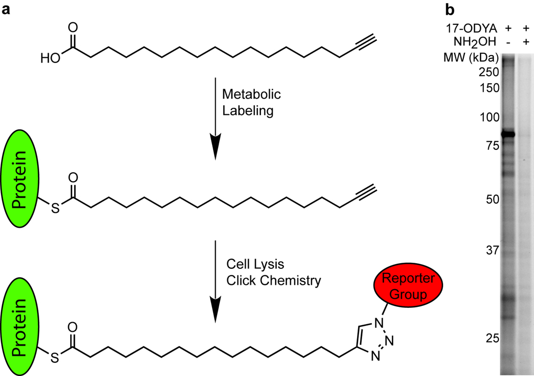
17-ODYA labeling and detection of palmitoylated proteins. (a) Schematic of 17-ODYA labeling. Cultured Jurkat T-cells were metabolically labeled with 17-ODYA and the lysates were then reacted with rhodamine-azide or biotin-azide for gel-based and LC-MS-based characterization of palmitoylated proteins. (b) Profiling palmitoylated proteins in the membrane fraction of Jurkat T-cells incubated with 25 µM 17-ODYA for 8 hours. Half of the sample was boiled in 2.5% hydroxylamine (NH2OH) for 5 minutes to hydrolyze thioesters and remove 17-ODYA labeling.
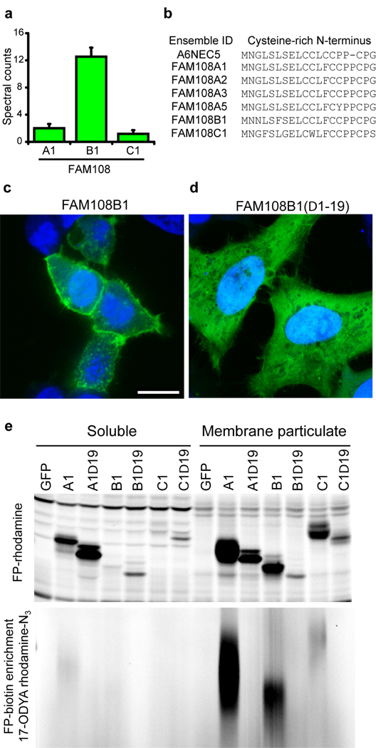
Membrane tethering of the FAM108 family of serine hydrolases by palmitoylation of an N-terminal cysteine-rich motif. (a)Average spectral counts of three FAM108 proteins identified by 17-ODYA profiling of Jurkat T-cells. None of these proteins showed any spectral counts in either the palmitate or hydroxylamine controls. (b) Cysteine-rich amino acid motif conserved among the seven members of the FAM108 family of proteins. FAM108A3 and FAM108A5 (which were not identified in this study) contain an additional N-terminal sequence preceding the cysteine-rich region of 73 and 28 amino acids respectively. (c) Distribution of FAM108B1-GFP fusion protein (green) expressed in HeLa cells co-stained with DAPI (blue). Scale bar = 15 µm. (d) Distribution of FAM108B1(Δ1–19)-GFP (green) expressed in HeLa cells co-stained with DAPI (blue). Deletion of the N-terminal cysteine-rich domain prevents FAM108B1 from localizing to the plasma membrane. (e) The N-terminal cysteine-rich motif of FAM108 proteins is palmitoylated and responsible FAM108 membrane localization. FAM108 proteins are fluorophosphonate (FP)-reactive serine hydrolases, which can be efficiently labeled with activity-based probes such as FP-rhodamine or FP-biotin. The upper panel shows FP-rhodamine-labeled soluble and membrane particulate fractions. The distribution of active FAM108 protein is altered from the particulate to soluble fractions by deletion of the N-terminal cysteine-rich motif. In the lower panel, 17-ODYA labeled transfected 293T cells were labeled with FP-biotin and enriched with streptavidin-agarose beads, reacted with rhodamine-azide, and visualized by in-gel fluorescence scanning.
References
Publication types
MeSH terms
Substances
Grants and funding
LinkOut - more resources
Full Text Sources
Other Literature Sources
