Divergent effects of PERK and IRE1 signaling on cell viability
- PMID: 19137072
- PMCID: PMC2614882
- DOI: 10.1371/journal.pone.0004170
Divergent effects of PERK and IRE1 signaling on cell viability
Abstract
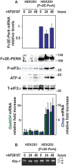
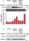
Protein misfolding in the endoplasmic reticulum (ER) activates a set of intracellular signaling pathways, collectively termed the Unfolded Protein Response (UPR). UPR signaling promotes cell survival by reducing misfolded protein levels. If homeostasis cannot be restored, UPR signaling promotes cell death. The molecular basis for the switch between prosurvival and proapoptotic UPR function is poorly understood. The ER-resident proteins, PERK and IRE1, control two key UPR signaling pathways. Protein misfolding concomitantly activates PERK and IRE1 and has clouded insight into their contributions toward life or death cell fates. Here, we employed chemical-genetic strategies to activate individually PERK or IRE1 uncoupled from protein misfolding. We found that sustained PERK signaling impaired cell proliferation and promoted apoptosis. By contrast, equivalent durations of IRE1 signaling enhanced cell proliferation without promoting cell death. These results demonstrate that extended PERK and IRE1 signaling have opposite effects on cell viability. Differential activation of PERK and IRE1 may determine life or death decisions after ER protein misfolding.
Conflict of interest statement
Figures


Similar articles
-
Imaging of single cell responses to ER stress indicates that the relative dynamics of IRE1/XBP1 and PERK/ATF4 signalling rather than a switch between signalling branches determine cell survival.Cell Death Differ. 2015 Sep;22(9):1502-16. doi: 10.1038/cdd.2014.241. Epub 2015 Jan 30. Cell Death Differ. 2015. PMID: 25633195 Free PMC article.
-
IRE1 signaling affects cell fate during the unfolded protein response.Science. 2007 Nov 9;318(5852):944-9. doi: 10.1126/science.1146361. Science. 2007. PMID: 17991856 Free PMC article.
-
Novel prosurvival function of Yip1A in human cervical cancer cells: constitutive activation of the IRE1 and PERK pathways of the unfolded protein response.Cell Death Dis. 2017 Mar 30;8(3):e2718. doi: 10.1038/cddis.2017.147. Cell Death Dis. 2017. PMID: 28358375 Free PMC article.
-
IRE1: ER stress sensor and cell fate executor.Trends Cell Biol. 2013 Nov;23(11):547-55. doi: 10.1016/j.tcb.2013.06.005. Epub 2013 Jul 21. Trends Cell Biol. 2013. PMID: 23880584 Free PMC article. Review.
-
Multiple Mechanisms of Unfolded Protein Response-Induced Cell Death.Am J Pathol. 2015 Jul;185(7):1800-8. doi: 10.1016/j.ajpath.2015.03.009. Epub 2015 May 5. Am J Pathol. 2015. PMID: 25956028 Free PMC article. Review.
Cited by
-
Remodeling the proteostasis network to rescue glucocerebrosidase variants by inhibiting ER-associated degradation and enhancing ER folding.PLoS One. 2013 Apr 19;8(4):e61418. doi: 10.1371/journal.pone.0061418. Print 2013. PLoS One. 2013. PMID: 23620750 Free PMC article.
-
Crizotinib and Doxorubicin Cooperatively Reduces Drug Resistance by Mitigating MDR1 to Increase Hepatocellular Carcinoma Cells Death.Front Oncol. 2021 May 18;11:650052. doi: 10.3389/fonc.2021.650052. eCollection 2021. Front Oncol. 2021. PMID: 34094940 Free PMC article.
-
An interdomain helix in IRE1α mediates the conformational change required for the sensor's activation.J Biol Chem. 2021 Jan-Jun;296:100781. doi: 10.1016/j.jbc.2021.100781. Epub 2021 May 14. J Biol Chem. 2021. PMID: 34000298 Free PMC article.
-
A unique modulator of endoplasmic reticulum stress-signalling pathways: the novel pharmacological properties of amiloride in glial cells.Br J Pharmacol. 2010 Jan 1;159(2):428-37. doi: 10.1111/j.1476-5381.2009.00544.x. Epub 2009 Dec 15. Br J Pharmacol. 2010. PMID: 20015086 Free PMC article.
-
Role of CAAT/enhancer binding protein homologous protein in panobinostat-mediated potentiation of bortezomib-induced lethal endoplasmic reticulum stress in mantle cell lymphoma cells.Clin Cancer Res. 2010 Oct 1;16(19):4742-54. doi: 10.1158/1078-0432.CCR-10-0529. Epub 2010 Jul 20. Clin Cancer Res. 2010. PMID: 20647473 Free PMC article.
References
-
- Ron D, Walter P. Signal integration in the endoplasmic reticulum unfolded protein response. Nat Rev Mol Cell Biol. 2007;8:519–529. - PubMed
-
- Harding HP, Zhang Y, Ron D. Protein translation and folding are coupled by an endoplasmic-reticulum-resident kinase. Nature. 1999;397:271–274. - PubMed
-
- Harding HP, Novoa I, Zhang Y, Zeng H, Wek R, et al. Regulated translation initiation controls stress-induced gene expression in mammalian cells. Mol Cell. 2000;6:1099–1108. - PubMed
-
- Zhou D, Palam LR, Jiang L, Narasimhan J, Staschke KA, et al. Phosphorylation of eIF2 directs ATF5 translational control in response to diverse stress conditions. J Biol Chem. 2008;283:7064–7073. - PubMed
-
- Calfon M, Zeng H, Urano F, Till JH, Hubbard SR, et al. IRE1 couples endoplasmic reticulum load to secretory capacity by processing the XBP-1 mRNA. Nature. 2002;415:92–96. - PubMed
Publication types
MeSH terms
Substances
Grants and funding
LinkOut - more resources
Full Text Sources
Other Literature Sources

