

Two- and three-dimensional models for risk assessment of radiation-enhanced colorectal tumorigenesis
- PMID: 19138051
- PMCID: PMC2659457
- DOI: 10.1667/RR1415.1
Two- and three-dimensional models for risk assessment of radiation-enhanced colorectal tumorigenesis
Abstract
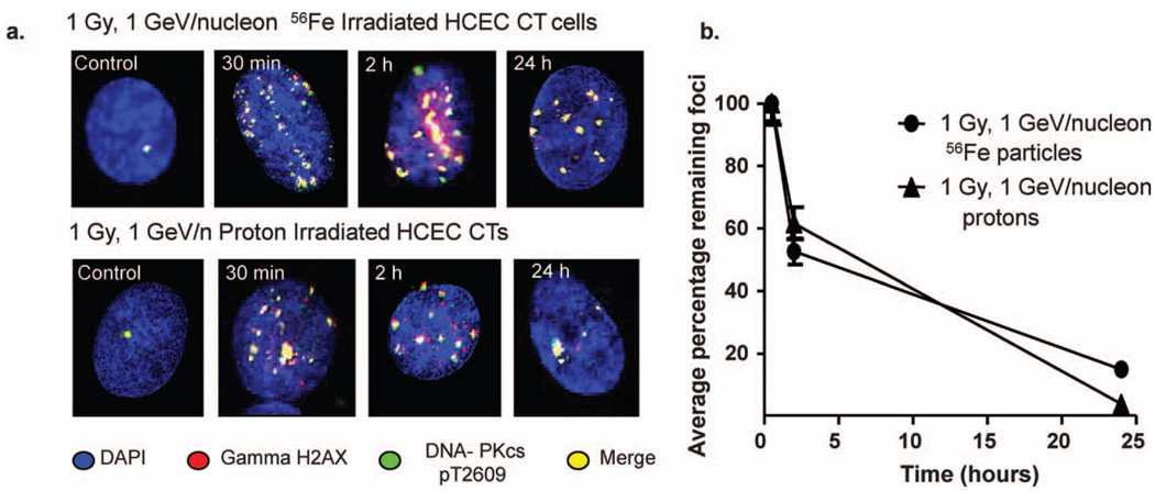
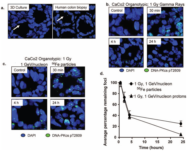
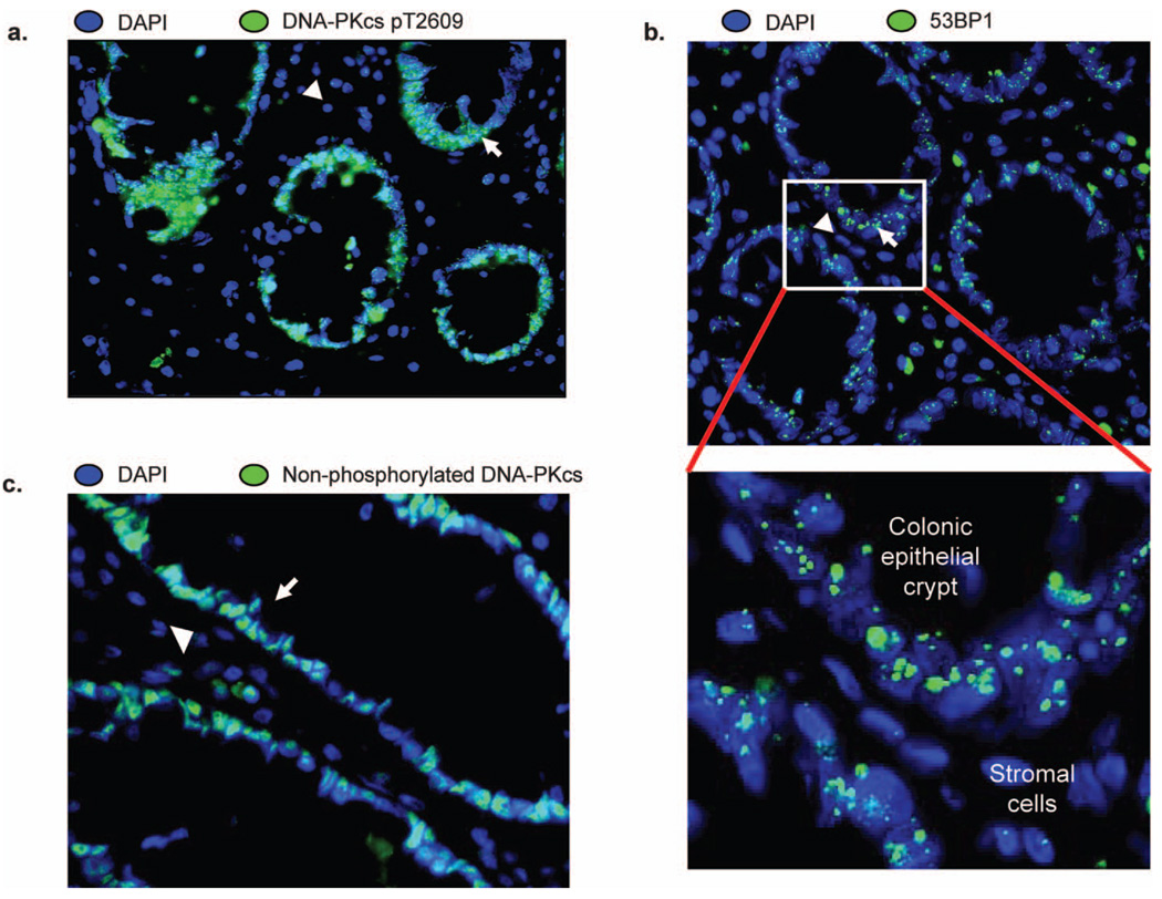
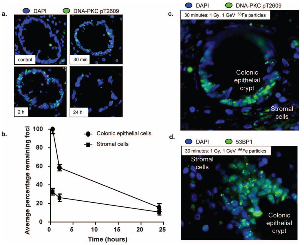
Astronauts may be at an increased risk for developing colorectal cancer after a prolonged interplanetary mission given the potential for greater carcinogenic effects of radiation to the colon. In addition, with an increase in age, there is a greater incidence of premalignant colon adenomas with age. In the present study, we have compared the effects of radiation on human colon epithelial cells in two-dimensional (2D) monolayer culture, in three-dimensional (3D) culture, and in intact human colon tissue biopsies. Immortalized colon epithelial cells were irradiated at the NASA Space Radiation Laboratory (NSRL) with either 1 Gy 1 GeV/nucleon (56)Fe particles or 1 Gy 1 GeV/nucleon protons and were stained at various times to assess DNA damage and repair responses. The results show more persisting damage at 24 h with iron-particle radiation compared to protons. Similar results were seen in 3D colon epithelial cell cultures in which (56)Fe-particle-irradiated specimens show more persisting damage at 24 h than those irradiated with low-LET gamma rays. We compared these results to those obtained from human colon tissue biopsies irradiated with 1 Gy gamma rays or 1 Gy 1 GeV (56)Fe particles. Observations of radiation-induced DNA damage and repair in gamma-irradiated specimens revealed more pronounced early DNA damage responses in the epithelial cell compartment compared to the stromal cell compartment. After low-LET irradiation, the damage foci mostly disappeared at 24 h. Antibodies to more than one type of DNA repair factor display this pattern of DNA damage, and staining of nonirradiated cells with nonphosphorylated DNA-PKcs shows a predominance of epithelial staining over stromal cells. Biopsy specimens irradiated with high-LET radiations also show a pattern of predominance of the DNA damage response in the highly proliferative epithelial cell compartment. Persistent unrepaired DNA damage in colon epithelial cells and the differing repair responses between the epithelial and mesenchymal compartments in tissues may enhance tumorigenesis by both stem cell transformation and alterations in the radiation-induced permissive tissue microenvironment that may potentiate cancer progression.
Figures
Similar articles
-
Response of thyroid follicular cells to gamma irradiation compared to proton irradiation. I. Initial characterization of DNA damage, micronucleus formation, apoptosis, cell survival, and cell cycle phase redistribution.Radiat Res. 2001 Jan;155(1 Pt 1):32-42. doi: 10.1667/0033-7587(2001)155[0032:rotfct]2.0.co;2. Radiat Res. 2001. PMID: 11121213
-
Relative effectiveness of HZE iron-56 particles for the induction of cytogenetic damage in vivo.Radiat Res. 2001 Feb;155(2):353-9. doi: 10.1667/0033-7587(2001)155[0353:reohip]2.0.co;2. Radiat Res. 2001. PMID: 11175671
-
Heavy ion radiation exposure triggered higher intestinal tumor frequency and greater β-catenin activation than γ radiation in APC(Min/+) mice.PLoS One. 2013;8(3):e59295. doi: 10.1371/journal.pone.0059295. Epub 2013 Mar 21. PLoS One. 2013. PMID: 23555653 Free PMC article.
-
Tissue responses to low protracted doses of high LET radiations or photons: early and late damage relevant to radio-protective countermeasures.Adv Space Res. 1989;9(10):299-313. doi: 10.1016/0273-1177(89)90453-5. Adv Space Res. 1989. PMID: 11537307 Review.
-
Understanding cancer development processes after HZE-particle exposure: roles of ROS, DNA damage repair and inflammation.Radiat Res. 2015 Jan;183(1):1-26. doi: 10.1667/RR13804.1. Epub 2015 Jan 7. Radiat Res. 2015. PMID: 25564719 Review.
Cited by
-
Exposure to heavy ion radiation induces persistent oxidative stress in mouse intestine.PLoS One. 2012;7(8):e42224. doi: 10.1371/journal.pone.0042224. Epub 2012 Aug 24. PLoS One. 2012. PMID: 22936983 Free PMC article.
-
High-LET-Radiation-Induced Persistent DNA Damage Response Signaling and Gastrointestinal Cancer Development.Curr Oncol. 2023 Jun 7;30(6):5497-5514. doi: 10.3390/curroncol30060416. Curr Oncol. 2023. PMID: 37366899 Free PMC article. Review.
-
Report of the AAPM TG-256 on the relative biological effectiveness of proton beams in radiation therapy.Med Phys. 2019 Mar;46(3):e53-e78. doi: 10.1002/mp.13390. Epub 2019 Feb 14. Med Phys. 2019. PMID: 30661238 Free PMC article.
-
Tissue slice cultures from humans or rodents: a new tool to evaluate biological effects of heavy ions.Radiat Environ Biophys. 2010 Aug;49(3):457-62. doi: 10.1007/s00411-010-0293-1. Epub 2010 May 19. Radiat Environ Biophys. 2010. PMID: 20490530
-
Truncated Adenomatous Polyposis Coli Mutation Induces Asef-Activated Golgi Fragmentation.Mol Cell Biol. 2018 Aug 15;38(17):e00135-18. doi: 10.1128/MCB.00135-18. Print 2018 Sep 1. Mol Cell Biol. 2018. PMID: 29866653 Free PMC article.
References
-
- Preston DL, Shimizu Y, Pierce DA, Suyama A, Mabuchi K. Studies of mortality of atomic bomb survivors. Report 13: Solid cancer and noncancer disease mortality: 1950–1997. Radiat. Res. 2003;160:381–407. - PubMed
-
- Cucinotta FA, Schimmerling W, Wilson JW, Peterson LE, Badhwar GD, Saganti PB, Dicello JF. Space radiation cancer risks and uncertainties for Mars missions. Radiat. Res. 2001;156:682–688. - PubMed
-
- Fearon ER, Vogelstein B. A genetic model for colorectal tumorigenesis. Cell. 1990;61:759–767. - PubMed
-
- Rex DK, Kahi CJ, Levin B, Smith RA, Bond JH, Brooks D, Burt RW, Byers T, Fletcher RH, Winawer SJ. Guidelines for colonoscopy surveillance after cancer resection: a consensus update by the American Cancer Society and the U.S. Multi-Society Task Force on Colorectal Cancer. Gastroenterology. 2006;130:1865–1871. - PubMed
-
- Rex DK, Lehman GA, Ulbright TM, Smith JJ, Pound DC, Hawes RH, Helper DJ, Wiersema MJ, Langefeld CD, Li W. Colonic neoplasia in asymptomatic persons with negative fecal occult blood tests: influence of age, gender, and family history. Am. J. Gastroenterol. 1993;88:825–831. - PubMed
Publication types
MeSH terms
Grants and funding
LinkOut - more resources
Full Text Sources
Medical
Molecular Biology Databases
