Review
. 2009 Jan;37(1):54-67.
doi: 10.1111/j.1442-9071.2008.01892.x. Epub 2008 Dec 9.

High-resolution ultrasound imaging of the eye - a review

Affiliations
Review

High-resolution ultrasound imaging of the eye - a review

Ronald H Silverman. Clin Exp Ophthalmol. 2009 Jan.

Abstract

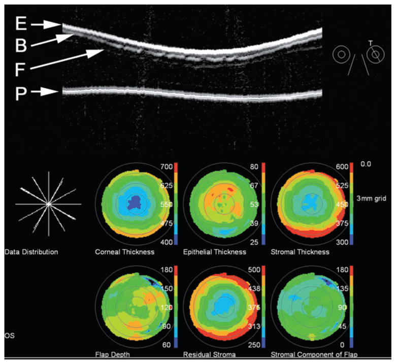
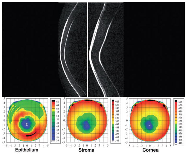
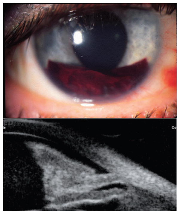
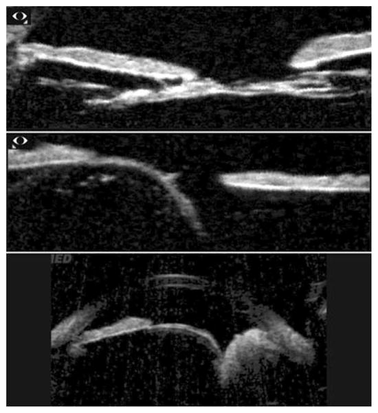
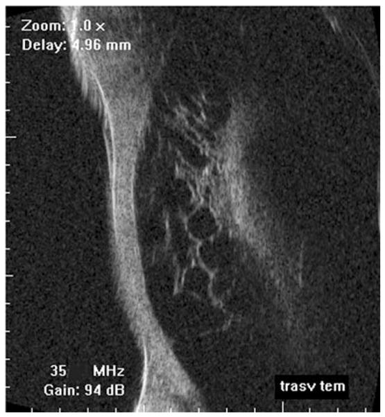
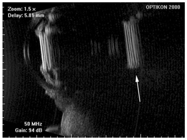
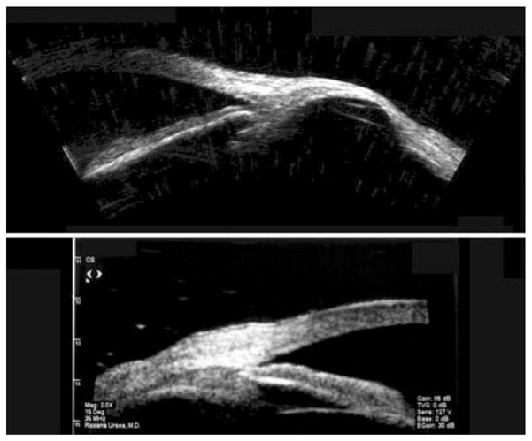
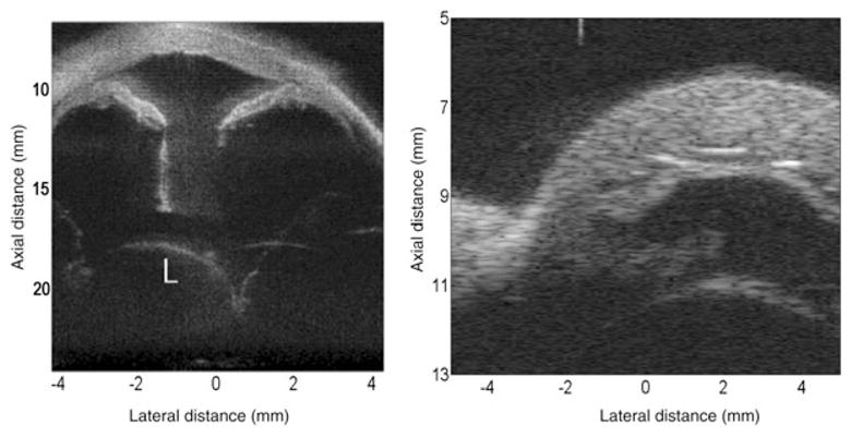
This report summarizes the physics, technology and clinical application of ultrasound biomicroscopy (UBM) of the eye, in which frequencies of 35 MHz and above provide over a threefold improvement in resolution compared with conventional ophthalmic ultrasound systems. UBM allows imaging of anatomy and pathology involving the anterior segment, including regions obscured by overlying optically opaque anatomic or pathologic structures. UBM provides diagnostically significant information in conditions such as glaucoma, cysts and neoplasms, trauma and foreign bodies. UBM also can provide crucial biometric information regarding anterior segment structures, including the cornea and its constituent layers and the anterior and posterior chambers. Although UBM has now been in use for over 15 years, new technologies, including transducer arrays, pulse encoding and combination of ultrasound with light, offer the potential for significant advances in high-resolution diagnostic imaging of the eye.


Figures

Figure 1
Figure 1
UBM image (Cornell prototype system) of normal anterior segment image acquired using arc-scan.
Figure 2
Figure 2
Top: Pupillary block is characterized by forward bowing of iris due to pressure differential between anterior and posterior chambers. Note cataract. Bottom: Plateau iris syndrome. Note characteristic forward position of ciliary processes and blunting of sulcus. Images acquired with Cornell arc-scan prototype system.
Figure 3
Figure 3
80 MHz image of angle structures temporally in eye 18 months post canaloplasty. Images acquired with IScience IUl-traSound system. (Image courtesy of Dr Hulgar Bull and Dr Kurt von Wolff.)
Figure 4
Figure 4
Retroiridal cysts may cause bowing of overlying iris, as shown here. Also note secondary cyst in ciliary body. Images acquired with Cornell arc-scan prototype system.
Figure 5
Figure 5
Top: Malignant melanoma of the iris at pupil margin. Middle: Pigmented lesion of the iris in the region of the angle demonstrating extension to the ciliary body. Bottom: Ciliary body melanoma. Note ‘cystic’ component, which may be attributable to regions within the melanoma differing in pigmentation and/or cell type. Images acquired with Cornell prototype arc-scanner.
Figure 6
Figure 6
Conjunctival spindle cell carcinoma. Top: Radial scan demonstrating a solid homogeneous conjunctival lesion, height: 3.04 mm. Bottom: Scan of adjacent area demonstrates extension into iridocorneal angle in anterior chamber. Scans performed with 50 MHz Sonomed VUMAX system. (Image courtesy of Norma Allemann, MD.)
Figure 7
Figure 7
Top: Artemis-2 B-mode images of corneal scar in horizontal plane in stretched rectilinear format (left) and geometrically corrected format (right). Bottom: Pachymetric maps formed from a series of scan meridians demonstrate scar location and extent. Corneal thickness map shows that the thinnest part of the cornea is displaced inferotemporally from the central cornea (thickness 514 μm) to the position of the scar, consistent with corneal thinning at the scar. The scar extends to a depth of approximately 300 mm, with an overall corneal thickness of about 470 mm at the scar position.
Figure 8
Figure 8
An incomplete flap occurred during the primary LASIK treatment of this eye because of the patient’s squeezing during the Hansatome microkeratome pass. Two months later, the patient was scanned using the Artemis-1 and the maximum flap thickness was measured to be 176 μm. The patient was then retreated using the VisuMax femtosecond laser (Carl Zeiss Meditec, Jena, Germany) with a programmed flap thickness of 190 μm so that the second flap would be deeper than the first flap. The images, top in geometrically correct format, bottom in stretched rectilinear format, show the cornea in the horizontal meridian after the second procedure. Both the initial, incomplete Hansatome flap and the complete VisuMax flap interfaces can be clearly seen. (Image courtesy of Dan Z. Reinstein, MD MA[Cantab] FRCSC.) LASIK, laser in situ keratomileusis.
Figure 9
Figure 9
Top: Artemis-2 scan of post-LASIK cornea shown in ‘stretched’ rectilinear format. The flap is readily seen. Also notable are interruptions in the Bowman’s membrane interface, suggestive of microfolds. Bottom: The pachymetric maps were reconstructed from measurements of the epithelial surface (E), Bowman’s membrane (B), the flap (F) and the posterior corneal surface (P) along scans at each of six clock-hours. Note irregular flap depth and thickening of the epithelium over the flap. LASIK, laser in situ keratomileusis.
Figure 10
Figure 10
Top: Geometrically and non-geometrically corrected vertical Artemis B-scan images of a patient with advanced keratoconus. Bottom: Pachymetric maps of the epithelium, stroma and cornea. Note area of epithelial thinning with a surround of epithelial thickening centred slightly inferior to the central cornea. This pattern occurs as the epithelium remodels in response to forward bulging of the underlying stroma. (Image courtesy of Dan Z. Reinstein, MD MA[Cantab] FRCSC.)
Figure 11
Figure 11
Hyphaema following bungee-cord injury, with organized blood inferiorly and more diffuse blood suspended in the anterior chamber. Scans performed with Sonomed UBM. (Image courtesy of Roxana Ursea, MD.)
Figure 12
Figure 12
Top: Congenital cataract demonstrates reduced lens thickness. Centre: Lens subluxation in Marfans’ syndrome. Bottom: Lens subluxation secondary to ciliary body tumour. Scans performed with Sonomed VUMAX. (Images courtesy of Norma Allemann, MD.)
Figure 13
Figure 13
Spontaneous ciliary body detachment. Scan performed by contact exam using probe cap on Optikon HiScan. (Image courtesy of Vincenzina Mazzeo, MD.)
Figure 14
Figure 14
Piggyback lens implants. Note reduplication artifact (arrow) resulting from multiple reflections of highly specular and reflective implants. Scans performed with Optikon HiScan. (Image courtesy of Vincenzina Mazzeo, MD.)
Figure 15
Figure 15
Top: Scleromalacia 6 years following ruptured globe. Image acquired using Cornell prototype scanner. Bottom: Thickened sclera in localized anterior uveitis, demonstrated with Sonomed UBM. (Image courtesy of Roxana Ursea, MD.)
Figure 16
Figure 16
Left: Image of anterior of ex vivo human eye made with 5-element 40 MHz annular array with chirp excitation. The combination of pulse encoding and synthetic focusing provides increased sensitivity and depth-of-field, enabling visualization of the dislocated lens. (Image courtesy of Jeffrey Ketterling, PhD and Jonathan Mamou, PhD.) Right: Anterior segment of ex vivo rabbit eye obtained with 64-element 30 MHz linear array using bipolar pulses and synthetic focusing. (Image courtesy of K. Kirk Shung, PhD, Jonathan Cannata, PhD and Xu Xiao-Chen, PhD.)

References

    1. Mundt GH, Hughes WF. Ultrasonics in ocular diagnosis. Am J Ophthalmol. 1956;42:488–98. - PubMed
    1. Baum G, Greenwood I. The application of ultrasonic locating techniques to ophthalmology. II. Ultrasonic slit-lamp in the ultrasonic visualization of soft tissues. Arch Ophthalmol. 1958;60:263–79. - PubMed
    1. Sherar MD, Foster FST. The design and fabrication of high frequency poly(vinylide fluoride) transducers. Ultrason Imag. 1989;11:75–94. - PubMed
    1. Foster FS, Harasiewicz KA, Sherar MD. A history of medical and biological imaging with polyvinylidene fluoride (PVDF) transducers. IEEE Trans Ultrason Ferroelectr Freq Control. 2000;47:1363–71. - PubMed
    1. Pavlin CJ, Sherar MD, Foster FS. Subsurface ultrasound microscopic imaging of the intact eye. Ophthalmology. 1990;97:244–50. - PubMed

Publication types

MeSH terms