. 2009 Jan 16;206(1):99-111.
doi: 10.1084/jem.20080521. Epub 2009 Jan 12.

Estrogen directly activates AID transcription and function

Affiliations

Estrogen directly activates AID transcription and function

Siim Pauklin et al. J Exp Med. .

Abstract

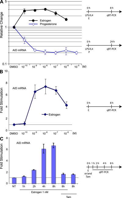
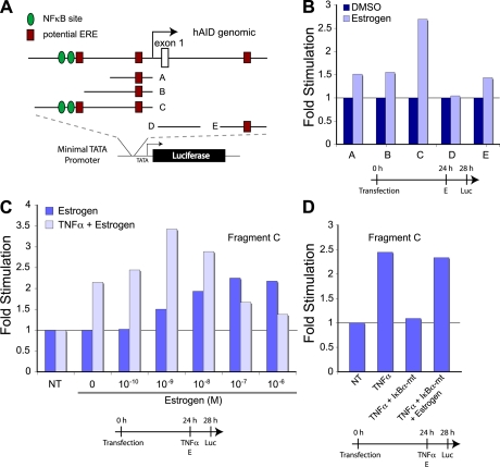
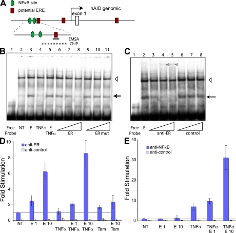
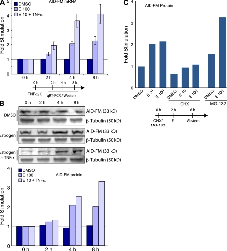
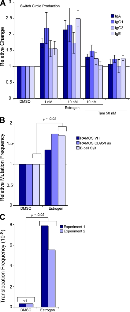
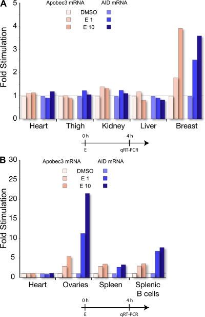
The immunological targets of estrogen at the molecular, humoral, and cellular level have been well documented, as has estrogen's role in establishing a gender bias in autoimmunity and cancer. During a healthy immune response, activation-induced deaminase (AID) deaminates cytosines at immunoglobulin (Ig) loci, initiating somatic hypermutation (SHM) and class switch recombination (CSR). Protein levels of nuclear AID are tightly controlled, as unregulated expression can lead to alterations in the immune response. Furthermore, hyperactivation of AID outside the immune system leads to oncogenesis. Here, we demonstrate that the estrogen-estrogen receptor complex binds to the AID promoter, enhancing AID messenger RNA expression, leading to a direct increase in AID protein production and alterations in SHM and CSR at the Ig locus. Enhanced translocations of the c-myc oncogene showed that the genotoxicity of estrogen via AID production was not limited to the Ig locus. Outside of the immune system (e.g., breast and ovaries), estrogen induced AID expression by >20-fold. The estrogen response was also partially conserved within the DNA deaminase family (APOBEC3B, -3F, and -3G), and could be inhibited by tamoxifen, an estrogen antagonist. We therefore suggest that estrogen-induced autoimmunity and oncogenesis may be derived through AID-dependent DNA instability.


Figures

Figure 1.
Figure 1.
The effects of estrogen and progesterone on AID mRNA in murine splenic B-cells. (A) AID mRNA in response to estrogen and progesterone treatment in stimulated B cells. Isolated mouse spleen B cells were stimulated with LPS and IL-4 and treated with different physiological concentrations of estrogen for 8 h or progesterone for 24 h. Unless indicated, DMSO is set to 1, and treatments are represented as relative change to DMSO. (B) AID mRNA in response to estrogen treatment in unstimulated B cells after 8 h treatment with physiological concentrations of estrogen. (C) AID mRNA induction upon different treatment. Cells were treated with 1 nM estrogen and/or 50 nM Tam (Tam) for up to 8 h. DMSO at 0 h is set to 1. All qRT-PCR data are representative of three independent experiments, and error bars indicate standard deviations from the mean. Timelines of cell treatments are indicated next to the graphs. NT, not treated. For A and B, absolute values as compared with GAPDH mRNA are shown in Fig. S10, available at http://www.jem.org/cgi/content/full/jem.20080521/DC1.
Figure 2.
Figure 2.
Human AID promoter analysis for hormone response elements. (A) Schematic representation of potential EREs (square) and NF-κB sites (circle) and their respective locations in the human promoter. The indicated promoter regions (marked A–E) were inserted into a luciferase reporter construct with a minimal promoter. The vectors were transfected into SiHa cells, incubated for 24 h, treated for 4 h with hormones or TNF-α, and analyzed for luciferase activity. (B) Relative luciferase activity after estrogen treatment. Cells were transfected with constructs containing AID promoter fragments and treated with estrogen for 4 h. (C) Effect of TNF-α and estrogen on the human AID promoter. Expression construct with an AID promoter region containing NF-κB sites and putative ERE (Fragment C) were transfected into cells, followed by TNF-α and/or estrogen treatment for 4 h. (D) Estrogen can act independently from NF-κB. Cells were cotransfected with Fragment C and an IκBα-mt expression vector. After 24 h, cells were treated with TNF-α and/or 100 nM estrogen for 4 h. Timelines of cell treatments are indicated below the graphs. NT, not treated.
Figure 3.
Figure 3.
Identification of ER binding to human AID promoter by EMSA and ChIP. (A) Schematic representation of human AID promoter region (as in Fig. 2 A). The position of the oligonucleotide used for EMSA and the region amplified by qRT-PCR for ChIP are marked as a black line and a dashed line, respectively. (B) Estrogen (denoted as E) -induced oligonucleotide shift (marked with an arrow) in Ramos nuclear extracts. Cells were treated for 72 h in hormone-depleted serum, followed by 4-h treatment with 10 nM estrogen (lanes 3 and 5–11) and/or TNF-α (lanes 4 and 5), and nuclear extract preparation. Different concentrations of unlabeled competitors ER (lanes 6–8) and mutated ER mut (lanes 9–11) were added to the binding reaction. Open triangle, nonspecific DNA binding band. (C) EMSA with anti-ERα antibodies. Increasing concentrations of anti-ERα antibody and a nonspecific antibody were added to the binding reaction (see Materials and methods). The estrogen-induced band is marked with an arrow, and a super-shifted band appearing upon anti-ERα antibody addition is marked with a closed triangle. Open triangle, nonspecific DNA-binding band. (D) ERα binds to upstream region of human AID promoter. Cells were treated as in B. Data are representative of three independent experiments and error bars indicate standard deviations from the mean. ChIP was performed using anti-ERα or control antibodies, and the bound DNA was subjected to qRT-PCR. Estrogen and Tam treatments are marked with E1 (estrogen 1 nM), E10 (estrogen 10 nM), and Tam, respectively. (E) Estrogen can cooperate with TNF-α in recruiting NF-κB to AID promoter. ChIP is as in D, using anti–NF-κB or control antibodies. NT, not treated.
Figure 4.
Figure 4.
The effects of estrogen on AID protein in DT40. (A) Estrogen induces AID mRNA expression. AID-FM–tagged DT40 cells were treated with DMSO, 100 nM estrogen, and 10 nM estrogen with TNF-α, lysed, and analyzed for AID mRNA expression with pRT-PCR at various time-points. (B) Estrogen induces AID-FM fusion protein expression. Treatment as in A, but lysates were analyzed by quantitative Western blot. For each sample, FLAG and Tubulin expression was quantitated. The graph is derived from correlating the FLAG expression to Tubulin expression, and then determining the ratio of estrogen-induced FLAG expression to untreated DMSO samples. (C) Estrogen does not affect AID-FM fusion protein stability. Cells were incubated with CHX or MG-132 for 2 h, followed by estrogen treatment for 4 h. Protein levels were determined by quantitative Western blot. For all experiments, cells were grown in hormone depleted media for 48 h. Results are normalized to control treatments as indicated on each graph. Timelines of cell treatments are indicated below the graphs.
Figure 5.
Figure 5.
Hormonal effects on Ig class switching, hypermutation, and translocation. (A) Estrogen induces isotype switching. Isolated mouse splenic B cells were stimulated for 48 h with LPS + IL-4 for switching to IgG1 and IgE, LPS + TGF-β for switching to IgA, and LPS for switching to IgG3. Indicated amounts of estrogen and/or Tam were added to the cells together with cytokines. Relative efficiency of class switching was determined by detecting circle transcripts with qRT-PCR, and data are normalized to the control treatment with DMSO from three independent experiments (error bars indicate standard deviations). (B) Estrogen increases the mutation frequency in VH and CD95/Fas loci of Ramos, and in Sγ3 of splenic mouse cells. Ramos cells were grown in the presence of 100 nM estrogen for ∼20 doublings, followed by sequencing of 341 bp from human VH or 750 bp from human CD95/Fas locus. Splenic mouse cells were treated for 6 d with LPS and 10 nM estrogen, and switch gamma3 loci amplified and sequenced (Fig. S7). Mutation frequencies are normalized to the control treatments with DMSO. A standard unpaired two-tailed Student's t test showed a significant difference in mutation frequency in the Sγ3 loci of DMSO- and estrogen-treated spleen cells. (C) Estrogen enhances the c-myc/IgH translocations in splenic B cells from p53+/− mice. In each experiment, 2 spleens per sample were treated with or without 50 nM estrogen in the presence of LPS for 72 h. More than 7 × 10⁷ cells were analyzed by long-range PCR (5 × 10⁴ cells/PCR; Fig. S8 and Supplemental materials and methods). Frequency was determined as c-myc/IgH translocation events per cell number analyzed. Statistics was performed on the results of the pooled experiments (two-tailed, unpaired Student's t test: P = 0.026). Fig. S7, Fig. S8, and Supplemental materials and methods are available at http://www.jem.org/cgi/content/full/jem.20080521/DC1.
Figure 6.
Figure 6.
Estrogen induces AID and Apobec3 transcription in mouse tissue. The red and blue colors indicate the results for mApobec3 and mAID, respectively. Tissues were treated with DMSO, 1 nM estrogen (E1), or 10 nM estrogen (E10). Gene expression is normalized to the control treatments with DMSO. The tissue expression profiles represent pooled data for the respective tissues from two experiments. Timelines of cell treatments are indicated below the graphs. Absolute values as compared with GAPDH mRNA are shown in Fig. S10, available at http://www.jem.org/cgi/content/full/jem.20080521/DC1.

Comment in

References

    1. Neuberger, M.S., R.S. Harris, J. Di Noia, and S.K. Petersen-Mahrt. 2003. Immunity through DNA deamination. Trends Biochem. Sci. 28:305–312. - PubMed
    1. Arakawa, H., J. Hauschild, and J.M. Buerstedde. 2002. Requirement of the activation-induced deaminase (AID) gene for immunoglobulin gene conversion. Science. 295:1301–1306. - PubMed
    1. Harris, R.S., J.E. Sale, S.K. Petersen-Mahrt, and M.S. Neuberger. 2002. AID is essential for immunoglobulin V gene conversion in a cultured B cell line. Curr. Biol. 12:435–438. - PubMed
    1. Muramatsu, M., K. Kinoshita, S. Fagarasan, S. Yamada, Y. Shinkai, and T. Honjo. 2000. Class switch recombination and hypermutation require activation-induced cytidine deaminase (AID), a potential RNA editing enzyme. Cell. 102:553–563. - PubMed
    1. Muramatsu, M., V.S. Sankaranand, S. Anant, M. Sugai, K. Kinoshita, N.O. Davidson, and T. Honjo. 1999. Specific expression of activation-induced cytidine deaminase (AID), a novel member of the RNA-editing deaminase family in germinal center B cells. J. Biol. Chem. 274:18470–18476. - PubMed

Publication types

MeSH terms