. 2009 Jan 16;206(1):209-19.
doi: 10.1084/jem.20082136. Epub 2009 Jan 12.

Natural micropolymorphism in human leukocyte antigens provides a basis for genetic control of antigen recognition

Affiliations

Natural micropolymorphism in human leukocyte antigens provides a basis for genetic control of antigen recognition

Julia K Archbold et al. J Exp Med. .

Abstract

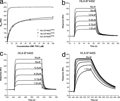
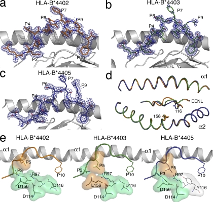
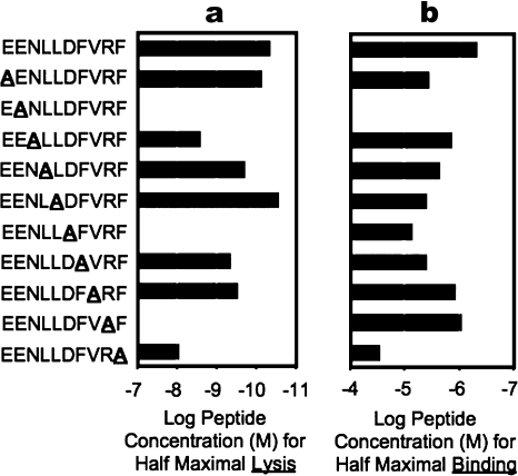
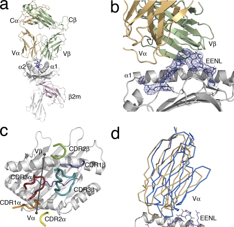
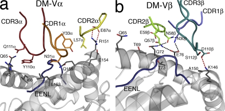
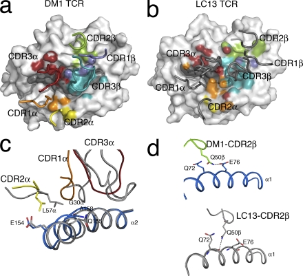
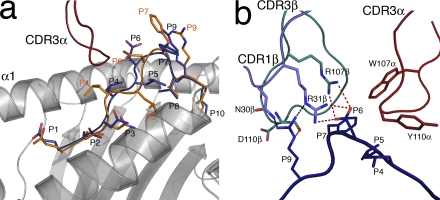
Human leukocyte antigen (HLA) gene polymorphism plays a critical role in protective immunity, disease susceptibility, autoimmunity, and drug hypersensitivity, yet the basis of how HLA polymorphism influences T cell receptor (TCR) recognition is unclear. We examined how a natural micropolymorphism in HLA-B44, an important and large HLA allelic family, affected antigen recognition. T cell-mediated immunity to an Epstein-Barr virus determinant (EENLLDFVRF) is enhanced when HLA-B*4405 was the presenting allotype compared with HLA-B*4402 or HLA-B*4403, each of which differ by just one amino acid. The micropolymorphism in these HLA-B44 allotypes altered the mode of binding and dynamics of the bound viral epitope. The structure of the TCR-HLA-B*4405(EENLLDFVRF) complex revealed that peptide flexibility was a critical parameter in enabling preferential engagement with HLA-B*4405 in comparison to HLA-B*4402/03. Accordingly, major histocompatibility complex (MHC) polymorphism can alter the dynamics of the peptide-MHC landscape, resulting in fine-tuning of T cell responses between closely related allotypes.


Figures

Figure 1.
Figure 1.
Differential affinity and binding kinetics of the DM1 TCR for the HLA-B44 allotypes. (a) Steady-state (or R equilibrium) binding of the three immobilized allotypes (HLA-B*4402EENL, HLA-B*4403EENL, and HLA-B*4405EENL) to the DM1 TCR. (b–d) Binding of increasing concentrations of the DM1 TCR (1.57–50 μM) to HLA-B*4402EENL (b), HLA-B*4403EENL (c), and HLA-B*4405EENL (d). Data are representative of at least four experiments. Kinetic association and dissociation rate constants were evaluated using the BIAevalution software. The DM1 TCR displayed a 10-fold higher affinity and a 10-fold slower dissociation rate constant for HLA-B*4405EENL over the other allotypes.
Figure 2.
Figure 2.
Structures of HLA-B*4402, HLA-B*4403, and HLA-B*4405 presenting EENLLDFVRF show marked differences in the centrally bulged region of the epitope. (a–c) Structure of the EENL peptide bound to (with associated 2Fo-Fc electron density contoured at 1σ) HLA-B*4402 (a), HLA-B*4403 (b), and HLA-B*4405 (c). The α2 helix has been removed for clarity. Potential TCR contact points are at P4, P6, P7, and P9. The central region (P5–P9) of the epitope showed mobility in all three allotypes. (d) An overlay of the three HLA-B44 allotypes (HLA-B*4402, orange; HLA-B*4403, green; HLA-B*4405, blue) revealing the minimal movement of the α helices and the movement of the central region (P5–P7) of the Cα backbones of the peptides. (e) The hydrophobic (orange) Leu at P5 of the epitope in HLA-B*4402 is pushed away from the unfavorable hydrophilic (green) patch composed of R97, D114, D116, and D156, resulting in movement of the peptide backbone toward the α1 helix. In HLA-B*4403 and HLA-B*4405, this patch is less hydrophilic because of either the hydrophobic Leu at position 156 or the partially hydrophobic Tyr at position 116 (white), and can accommodate the hydrophobic P5 residue.
Figure 3.
Figure 3.
Alanine scanning mutagenesis of the peptide reveals a C-terminal focus. The impact of single amino-acid substitutions within the EENLLDFVRF peptide upon CTL recognition and HLA-B*4405 binding revealed P6 and P9 to be critical for TCR recognition. (a) EENL-specific CTL clones were tested for recognition of a panel of altered peptide ligands in which a single alanine substitution was introduced at each peptide position. A range of peptide concentrations was used in the chromium release assays, and the concentration required for half-maximum lysis was determined from the dose–response data. (b) MHC peptide-binding assays were also conducted at a range of concentrations for their ability to stabilize HLA-B*4405 expression. The concentration of peptide required for half-maximum HLA-B*4405 stabilization was calculated. Experiments were conducted once, with each sample tested in duplicate.
Figure 4.
Figure 4.
Overview of the DM1 TCR in complex with HLA-B*4405EENL. (a) The DM1–HLA-B*4405EENL complex (DM1 TCR Vα, orange; DM1 TCR Vβ, green; HLA-B*4405, gray; EENL peptide, blue; β2m, pink). (b) A closer view of the DM1 TCR binding to the HLA-B*4405EENL, showing the clear electron density for the peptide (2Fo-Fc map contoured at 1σ). (c) Relative docking angle of the DM1 TCR on the pMHC (80°). For docking orientation, an axis was drawn through the points at the center of mass of the Vα and Vβ domains. The positioning of the CDR loops of the DM1 TCR on HLA-B*4405 is also shown (CDR1α, orange; CDR2α, yellow; CDR3α, red; CDR1β, slate; CDR2β green; CDR3β, teal). (d) Scissoring action of the Vα domain compared with the Vβ domain upon ligation. Superposition of the Vβ domains of the nonliganded and liganded DM1 TCR revealed movement of the Vα domain by 6°. The nonliganded Vα domain of the TCR is shown in blue, and the liganded Vα domain of the TCR is shown in orange.
Figure 5.
Figure 5.
DM1 interactions with HLA-B*4405EENL. (a) Interactions with the HLA-B*4405 heavy chain mediated by the Vα domain of the DM1 TCR (CDR1α, orange; CDR2α, yellow; CDR3α, red; HLA-B*4405 heavy chain, gray; EENL epitope, blue). (b) Interactions with the HLA-B*4405 heavy chain mediated by the Vβ domain of the DM1 TCR (CDR1β, slate; CDR2β, green; CDR3β, teal; HLA-B*4405 heavy chain, gray; EENL epitope, blue).
Figure 6.
Figure 6.
DM1–pMHC interactions compared with the LC13–pMHC structure, revealing similar positioning of the CDRα loops. (a) DM1 TCR “footprint” on HLA-B*4405EENL. HLA-B*4405EENL (gray) is shown in surface representation. The pMHC residues are colored according to the relevant contacting CDR loop. The positions of the CDR loops are also shown (CDR1α, orange; CDR2α, yellow; CDR3α, red; CDR1β, slate; CDR2β, green; CDR3β, teal). (b) LC13 footprint on HLA-B*0801FLR. HLA-B*0801FLR (gray) is shown in surface representation. The pMHC residues are colored according to the relevant contacting CDR loop (CDR1α, orange; CDR2α, yellow; CDR3α, red; CDR1β, slate; CDR2β, green; CDR3β, teal). The LC13 CDR loops are shown in gray. (c) Similar positioning of the CDR1α loops (CDR1α, orange; CDR2α, yellow; CDR3α, red) of DM1 compared with LC13 (gray) allowed conservation of interactions with residues E154, Q155, and A158 on the α2 helix. (d) Although the positioning of the Vβ loops of the two TCRs was divergent, the packing of Q50β of the CDR2β loop of DM1 (green) and LC13 (gray) between Q72 and E76 was maintained.
Figure 7.
Figure 7.
DM1-mediated peptide interactions. (a) Conformational movement of the EENL peptide upon ligation by the DM1 TCR revealing plasticity of the pMHC surface. The nonligand-bound conformation is shown in orange, and the ligand-bound conformation of the EENL epitope is shown in blue. (b) DM1 interactions with the EENL peptide. A cluster of charged interactions between the DM1 TCR and the peptide provides specificity. This specificity is driven by CDR3α (red), CDR1β (slate), and CDR3β (teal). Salt bridges are shown as red dashed lines, and H bonds are shown as black dashed lines.

References

    1. Messaoudi, I., J.A. Patino, R. Dyall, J. LeMaoult, and J. Nikolich-Zugich. 2002. Direct link between MHC polymorphism, T cell avidity, and diversity in immune defense. Science. 298:1797–1800. - PubMed
    1. DiBrino, M., K.C. Parker, D.H. Margulies, J. Shiloach, R.V. Turner, W.E. Biddison, and J.E. Coligan. 1995. Identification of the peptide binding motif for HLA-B44, one of the most common HLA-B alleles in the Caucasian population. Biochemistry. 34:10130–10138. - PubMed
    1. Godfrey, D.I., J. Rossjohn, and J. McCluskey. 2008. The fidelity, occasional promiscuity, and versatility of T cell receptor recognition. Immunity. 28:304–314. - PubMed
    1. Clements, C.S., M.A. Dunstone, W.A. Macdonald, J. McCluskey, and J. Rossjohn. 2006. Specificity on a knife-edge: the alphabeta T cell receptor. Curr. Opin. Struct. Biol. 16:787–795. - PubMed
    1. Rudolph, M.G., R.L. Stanfield, and I.A. Wilson. 2006. How TCRs bind MHCs, peptides, and coreceptors. Annu. Rev. Immunol. 24:419–466. - PubMed

Publication types

MeSH terms