. 2009 Jan 12;184(1):113-27.
doi: 10.1083/jcb.200806044.

The SH3 domains of two PCH family members cooperate in assembly of the Schizosaccharomyces pombe contractile ring

Affiliations

The SH3 domains of two PCH family members cooperate in assembly of the Schizosaccharomyces pombe contractile ring

Rachel H Roberts-Galbraith et al. J Cell Biol. .

Abstract

Schizosaccharomyces pombe cdc15 homology (PCH) family members participate in many cellular processes by bridging the plasma membrane and cytoskeleton. Their F-BAR domains bind and curve membranes, whereas other domains, typically SH3 domains, are expected to provide cytoskeletal links. We tested this prevailing model of functional division in the founding member of the family, Cdc15, which is essential for cytokinesis in S. pombe, and in the related PCH protein, Imp2. We find that the distinct functions of Imp2 and Cdc15 are SH3 domain independent. However, the Cdc15 and Imp2 SH3 domains share an essential role in recruiting proteins to the contractile ring, including Pxl1 and Fic1. Together, Pxl1 and Fic1, a previously uncharacterized C2 domain protein, add structural integrity to the contractile ring and prevent it from fragmenting during division. Our data indicate that the F-BAR proteins Cdc15 and Imp2 contribute to a single biological process with both distinct and overlapping functions.


Figures

Figure 1.
Figure 1.
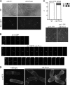
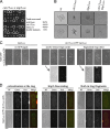
cdc15ΔSH3 cells are viable but exhibit cytokinesis defects. (A) cdc15ΔSH3 cells were grown to midlog phase, fixed, and stained with DAPI and methyl blue to visualize DNA and cell walls, respectively. The asterisk indicates a cell with both cell separation and cytokinesis defects, the arrow marks a “paired” cell, the arrowhead denotes a cell with impaired cell separation, and the double arrowhead marks a cell with a “kissing nuclei” phenotype. The boxed area is from a separate micrograph but was included to demonstrate all of the phenotypes scored. (B) Denatured lysates were prepared from wild-type, cdc15ΔSH3, and cdc15ΔSH3-FLAG3 cells grown at 32°C and immunoblotted with anti-Cdc15 serum or anti–α-tubulin for loading control. (C) Wild-type, cdc15ΔSH3-FLAG3, and cdc15ΔSH3 cells were grown at the temperatures indicated, fixed, and imaged as in A (n = 200). (D) cdc15-GFP and cdc15ΔSH3-GFP cells were grown to midlog phase at 25°C and imaged. Arrows indicate interphase cells, asterisks indicate contractile rings that have not begun constriction, and arrowheads mark constricting rings. The insets show a cell in which nodes of Cdc15ΔSH3 are present. BF, bright field. Bars: (A) 10 μm; (D) 5 μm.
Figure 2.
Figure 2.
cdc15ΔSH3 cells show defects in contractile ring dynamics. (A) rlc1-GFP and rlc1-GFP cdc15ΔSH3-FLAG3 cells were grown and imaged as in Fig. 1 D. BF, bright field. (B) rlc1-GFP and cdc15ΔSH3-FLAG3 rlc1-GFP cells were imaged live at 25°C every 2.5 min (Videos 1 and 2, available at http://www.jcb.org/cgi/content/full/jcb.200806044/DC1). Montages of cells completing cytokinesis are shown. Each image is 12.5 min apart. (C) cps1-191 rlc1-GFP and cps1-191 cdc15ΔSH3-FLAG3 rlc1-GFP cells were arrested and imaged. In the images, cells were divided into fifths, and the location of the contractile ring was quantified (n = 100). wt, wild type. (D) acyl-GFP was expressed in wild-type or cdc15ΔSH3-FLAG3 cells and imaged live. Membrane bridges remaining after cytokinesis are marked with arrows. Bars: (A and C) 5 μm; (B) 2 μm; (D) 3 μm.
Figure 3.
Figure 3.
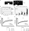
Dynamics of rings and ring components in cdc15ΔSH3. (A) Kymographs were created from lines drawn across the division site in five videos for each rlc1-GFP and cdc15ΔSH3-FLAG3 rlc1-GFP. Time is depicted on the vertical scale bar. (B) Contractile ring event timing for each cell in A was determined and averaged (n = 5 each; Fig. S1 H, available at http://www.jcb.org/cgi/content/full/jcb.200806044/DC1). Timing of cytokinesis in wild-type (wt) cells was plotted on the y axis, and the time taken for corresponding events in wild-type and cdc15ΔSH3-FLAG3 cells was plotted on the x axis. (C) The time taken for wild-type and cdc15ΔSH3-FLAG3 cells to complete stages of cytokinesis was determined for each genotype, and differences were determined by Student's t tests. P-values and SEM (error bars) are included. (D) FRAP measurements of Cdc15-GFP or Cdc15ΔSH3-GFP signals are shown (n = 24 each). t1/2 and mobile fraction values were calculated from best-fit curves and are shown below. The difference in t1/2 is significant (P = 0.046), as is the difference in Fm (P = 0.0009). R2 values for wild-type and cdc15ΔSH3 curves are 0.9753 and 0.9752, respectively. (E) FRAP measurements of Rlc1-GFP in cdc15+ or cdc15ΔSH3-FLAG3 backgrounds (n = 28 each). t1/2 and mobile fraction values were calculated from best-fit curves. The differences in t1/2 and Fm are not significant (P = 0.651 and P = 0.589, respectively). R2 values for wild-type and cdc15ΔSH3 curves are 0.9365 and 0.9518, respectively.
Figure 4.
Figure 4.
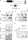
Cdc15 SH3 domain is necessary and sufficient for interaction with C2 domain protein Fic1. (A) Schematic representations of the domains of Cdc15 (top) and Fic1 (bottom). Domains are as indicated, except the PEST domain of Cdc15 is the white oval, and PXXP motifs in Fic1 are indicated with asterisks. Fragments of cdc15 and fic1 from the two-hybrid screen are depicted. (B and C) Coimmunoprecipitation of endogenously tagged Fic1-FLAG3 and Cdc15-HA3 from asynchronous cell lysate. Asterisks indicate a background band present in FLAG immunoblots. (D) Coimmunoprecipitations were performed in which Cdc15 or Cdc15ΔSH3 was isolated using anti-Cdc15 serum, or Fic1-FLAG3 was isolated with the anti-FLAG antibody, and bound proteins were identified by immunoblotting with either the anti-FLAG antibody or anti-Cdc15 serum. (E) Recombinant MBP or MBP-Cdc15SH3 was purified and incubated with recombinant bead-bound His6 or Fic1-His6. Beads were washed, and bound proteins were run on SDS-PAGE and detected by Coomassie staining. One tenth of each MBP protein input is shown as a control. IP, immunoprecipitation; WB, Western blot.
Figure 5.
Figure 5.
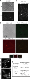
Fic1 localizes to the contractile ring and plays a nonessential role in cytokinesis. (A and B) fic1-GFP cells were imaged live. In B, one 0.5-μm slice was taken through cell middles to allow imaging of fainter structures. BF, bright field. (C) cdc15-CFP fic1-YFP cells were imaged through one 0.5-μm plane. Bright field, CFP (green), YFP (red), and merged images are shown. Insets (boxed area) show colocalization of Cdc15 and Fic1 at cell tips (arrows). (D) Wild-type and fic1Δ cells were stained as in Fig. 1 A. Quantitation of cell phenotypes is shown in Fig. S3 E (available at http://www.jcb.org/cgi/content/full/jcb.200806044/DC1). (E) fic1Δ was crossed with several mutants or deletions in cytokinesis genes to determine synthetic interactions. Bars: (A–D) 5 μm; (C, inset) 2 μm.
Figure 6.
Figure 6.
Fic1 binds the SH3 domain of PCH family member Imp2. (A) Coimmunoprecipitations of Imp2-HA3 and Fic1-FLAG3 were performed as in Fig. 4 (B and C). IP, immunoprecipitation. (B) Fic1-TAP–containing complexes were purified from nda3-km311 imp2-HA3 cells using IgG beads followed by TEV cleavage. These complexes were immunoprecipitated using either an anti-HA antibody or anti-Cdc15 serum, and bound proteins were detected by immunoblotting. WB, Western blot. (C) Lysates from wild-type, imp2-FLAG3, or imp2ΔSH3-FLAG3 cells were incubated with recombinant bead-bound His6 or Fic1-His6. Bound proteins were detected by immunoblotting. (D) An in vitro binding experiment was performed with MBP-Imp2SH3 and Fic1-His6 as in Fig. 4 E. (E) Wild-type, imp2ΔSH3-HA3, and imp2Δ cells were fixed and imaged as in Fig. 1 A (quantitation in Fig. S4 D, available at http://www.jcb.org/cgi/content/full/jcb.200806044/DC1). (F) Imp2ΔSH3-GFP was visualized in live cells. BF, bright field. Bars: (E) 10 μm; (F) 5 μm.
Figure 7.
Figure 7.
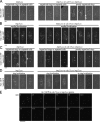
SH3 domains of Cdc15 and Imp2 function cooperatively in cytokinesis. (A) cdc15ΔSH3-FLAG–KanR and imp2ΔSH3-FLAG–HygR cells were crossed, and tetrads were pulled. Circles indicate missing double mutants, and the recovery rates are listed. (B) Images were taken for several resulting tetrads, and single colonies of each genotype are shown. wt, wild type. (C) Spores of the indicated genotypes were germinated overnight and imaged in the first cell division. Bright field images are overlaid onto GFP images. Below, bright field (BF) and GFP images of two germinating spores before (left) and after (right) initiation of septation are shown separately. (D) Spores were germinated as in C and imaged for both Imp2ΔSH3-GFP and Cdc15ΔSH3-mCherry localization. Bars, 2 μm.
Figure 8.
Figure 8.
Contractile ring defects of cdc15ΔSH3 imp2ΔSH3 spores. (A–D) Spores of the indicated genotypes were germinated in the presence of hygromycin overnight and imaged as in Fig. 7 C. (E) Images were taken at 150-s intervals from a video of Rlc1-GFP and Sid4-GFP in the cdc15ΔSH3 imp2ΔSH3 background. Time on the montage is indicated in minutes. Bars: (A–D), 2 μm; (E) 5 μm.
Figure 9.
Figure 9.
Pxl1 binds the SH3 domain of Cdc15 and functions in parallel with Fic1. (A) Diagram of Pxl1 to scale. LIM domains are shown as gray ovals, and PXXP motifs are depicted as asterisks. The site of the truncation used in the in vitro binding assay is indicated with an arrow. (B) Coimmunoprecipitation of Cdc15 with HA-Pxl1 was performed as in Fig. 4 D. The asterisk indicates a background band, and the arrow marks the HA-Pxl1 bands. IP, immunoprecipitation; WB, Western blot. (C) in vitro binding assay was performed as in Fig. 4 E. Binding is indicated with an asterisk. (D) GFP-pxl1 and cdc15ΔSH3 GFP-pxl1 strains were grown at 25°C and imaged live. (E) Spores were germinated and imaged as in Fig. 8 (A–D). BF, bright field. Bars, 5 μm.
Figure 10.
Figure 10.
The Imp2 SH3 domain can fulfill most functions of the Cdc15 SH3 domain. (A) The SH3 domain of Cdc15 was replaced with that of Imp2 at the endogenous cdc15 locus. Cells were grown and stained as in Fig. 1 A. (B) Rlc1-GFP and Sid4-GFP were imaged live in wild-type (wt) and cdc15-imp2SH3 backgrounds. BF, bright field. (C) Cdc15 and Imp2 are depicted as dimers at the membrane; F-BAR domains are shown as crescents, and SH3 domains are shown as dark ovals. In wild-type cells, both Cdc15 and Imp2 bind Fic1 (the C2 domain–containing protein) and possibly other proteins (Y). Cdc15 binds to Pxl1 as well. In the absence of the SH3 domain of Cdc15, some proteins (Fic1, Pxl1, and Y) continue to localize to the contractile ring through Imp2, whereas other Cdc15-specific proteins (X) fail to localize. In the absence of the SH3 domains of either Imp2 or Cdc15, none of these proteins localize properly, resulting in cytokinetic failure. Bars, 5 μm.

Similar articles

Cited by

References

    1. Arai, R., and I. Mabuchi. 2002. F-actin ring formation and the role of F-actin cables in the fission yeast Schizosaccharomyces pombe. J. Cell Sci. 115:887–898. - PubMed
    1. Bahler, J., J.Q. Wu, M.S. Longtine, N.G. Shah, A. McKenzie III, A.B. Steever, A. Wach, P. Philippsen, and J.R. Pringle. 1998. Heterologous modules for efficient and versatile PCR-based gene targeting in Schizosaccharomyces pombe. Yeast. 14:943–951. - PubMed
    1. Barr, F.A., and U. Gruneberg. 2007. Cytokinesis: placing and making the final cut. Cell. 131:847–860. - PubMed
    1. Carnahan, R.H., and K.L. Gould. 2003. The PCH family protein, Cdc15p, recruits two F-actin nucleation pathways to coordinate cytokinetic actin ring formation in Schizosaccharomyces pombe. J. Cell Biol. 162:851–862. - PMC - PubMed
    1. Celton-Morizur, S., N. Bordes, V. Fraisier, P.T. Tran, and A. Paoletti. 2004. C-terminal anchoring of mid1p to membranes stabilizes cytokinetic ring position in early mitosis in fission yeast. Mol. Cell. Biol. 24:10621–10635. - PMC - PubMed

Publication types

MeSH terms