. 2009 Apr;29(7):1944-58.
doi: 10.1128/MCB.00840-08. Epub 2009 Jan 12.

SALL3 interacts with DNMT3A and shows the ability to inhibit CpG island methylation in hepatocellular carcinoma

Affiliations

SALL3 interacts with DNMT3A and shows the ability to inhibit CpG island methylation in hepatocellular carcinoma

Yuko Shikauchi et al. Mol Cell Biol. 2009 Apr.

Abstract

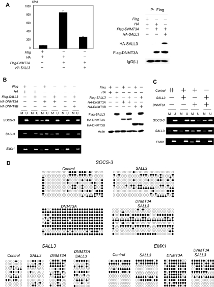
The mechanisms of aberrant CpG island methylation in oncogenesis are not fully characterized. In particular, little is known about the mechanisms of inhibition of CpG island methylation. Here we show that sal-like 3 (SALL3) is a novel inhibitory factor for DNA methyltransferase 3 alpha (DNMT3A). SALL3 binds to DNMT3A by a direct interaction between the double zinc finger motif of SALL3 and the PWWP domain of DNMT3A. SALL3 expression reduces DNMT3A-mediated CpG island methylation in cell culture and in vitro. CpG island methylation is enhanced in SALL3-depleted cells. Consistently, DNMT3A from SALL3-depleted cells increases methyltransferase activity in vitro. Binding of DNMT3A to chromatin is reduced or increased by SALL3 expression or depletion, respectively, accounting for the mechanism by which SALL3 inhibits DNMT3A-mediated CpG island methylation. We also show that SALL3 is inducible by BMP-4 and silenced by associated DNA methylation in hepatocellular carcinoma (HCC). Our results suggest that silencing of SALL3 results in acceleration of DNA methylation in HCC. This functional characterization of SALL3 sheds light on regulatory mechanisms for DNMT3A and provides new strategies to inhibit aberrant methylation in cancer.


Figures

FIG. 1.
FIG. 1.
Methylation-associated silencing of SALL3. (A) Schematic representation of the SALL3c protein. SALL3c contains eight C2H2-type zinc fingers (vertical bars). Four fingers compose two DZF motifs. NP_741996 and Q9BXA9 are coded by NM_19999 and AJ007421, respectively. (B) Upper panel, CpG density in the SALL3 promoter region. Vertical bars indicate CpG sequences. The translation start site is indicated as “1.” Arrows and arrowheads represent primer sets for MSP and bisulfite sequencing, respectively. Bottom panel, MSP analysis of primary HCC cell lines. HuH1 through FLC4 are HCC cell lines. Visible bands in lanes M are methylated products, and those in lanes U are unmethylated products. (C) Bisulfite sequencing analysis. Methylation status of the 5′ region of SALL3 (from −3139 through −2431) was investigated in three HCC cell lines (FLC4, HuH2, and Hep3B) and a normal liver sample. Ten individual clones were sequenced for each sample. Filled and open circles represent methylation and unmethylation, respectively. (D) RT-PCR analysis. Total RNA samples from 10 HCC cell lines and a normal liver were analyzed by RT-PCR with SALL3-specific primers (upper panel). GAPDH amplification verified the consistency of the RT-PCR (bottom panel). (E) Reactivation by a methylation inhibitor. Three methylated cell lines (Hep3B, HuH2, and HuH7) were treated with or without 5Aza-dC, and SALL3 expression was analyzed by RT-PCR. (F) MSP of primary HCC samples. Methylation of SALL3 was analyzed in 30 primary HCC samples by MSP. Representatives from the 30 samples are shown. Tumor samples are methylated in cases 1 through 5 and unmethylated in cases 6 through 10. “T” represents a tumor sample, and “N” represents its nontumorous counterpart. (G) Colony formation assay. Methylation-silenced cells (Hep3B and HuH2) were transfected with either the SALL3 expression vector or the backbone vector and selected for 4 weeks with G418.
FIG. 2.
FIG. 2.
Interaction of SALL3 with DNMT3. (A) Binding of FL-SALL3 to DNMT3. HA-SALL3 or the backbone vector (HA) was cotransfected with the Flag-DNMT3A or Flag-DNMT3B expression vector. Anti-HA immunoprecipitates were analyzed by immunoblotting with anti-Flag and anti-HA antibodies. (B) Lysate from nontransfected HEK293 cells was immunoprecipitated (IP) with anti-DNMT3A antibody or normal rabbit IgG. The immunoblot was analyzed with anti-SALL3 and anti-DNMT3A antibodies. IVT is in vitro-transcribed-translated SALL3. IgG was used as a control to show that the anti-DNMT3A antibody does not precipitate SALL3 nonspecifically (left panel). Anti-DNMT3A immunoprecipitation was performed using HEK293 cells transfected with DNMT3A-specific siRNA or control siRNA (right panel). (C) HA-tagged SALL3 or HA-stably expressing cells were generated by transfection of HA-SALL3 or HA tag alone, respectively, and subsequent selection with G418. The cell lysates were immunoprecipitated with anti-HA antibody and analyzed by immunoblotting with anti-DNMT3A and anti-HA antibodies. (D) Interaction of SALL3 deletion mutants containing the DZF motif with DNMT3A. Various Flag-tagged SALL3 deletion mutants were cotransfected with FL-DNMT3A. Anti-Flag immunoprecipitates were analyzed by immunoblotting with anti-DNMT3A antibody. Deletions 1 through 6 are shown schematically (left panel). The cell lysates were analyzed by immunoblotting with anti-DNMT3A and anti-Flag antibodies (right panel). Vector is the vector control. FL is FL-SALL3. (E) Direct binding of SALL3 deletion mutants containing the DZF motif to DNMT3A in vitro. Immobilized GST-FL-DNMT3A or GST alone was mixed with in vitro-translated 35S-labeled FL-SALL3 or SALL3 deletion mutants (1, 3, or 6). Beads were washed, and bound proteins were analyzed by SDS-PAGE and autoradiography. (F) Direct binding in vitro of DNMT3A deletion mutants containing the PWWP domain to the SALL3 deletion mutant containing the DZF motif. A GST pull-down assay was performed using immobilized GST-SALL3 deletion mutant 6 and in vitro-translated 35S-labeled FL-DNMT3A or DNMT3A deletion mutants (schematically shown in the upper panel).
FIG. 3.
FIG. 3.
DNMT3A-mediated CpG island methylation and its inhibition by SALL3. (A) In vitro methyltransferase activity assay. DNMT3A was isolated by anti-Flag immunoprecipitations from cells transfected with the plasmids indicated. A methyltransferase reaction was carried out using isolated DNMT3A, S-adenosyl-l-[methyl-3H]methionine, and SOCS-3-plasmid DNA. The values are given as means ± standard deviations of results for the three replicates. The difference in DNMT3A activity between cells transfected with DNMT3A and those transfected with DNMT3A and SALL3 is statistically significant (P = 2.0E−05) by a Student t test (left panel). The anti-Flag immunoprecipitates were analyzed by immunoblotting with anti-Flag and anti-HA antibodies (right panel). (B) MSP analysis of transfected clone 12 cells in SOCS-3, SALL3, and EMX1 CpG islands. The cells were transiently transfected with the plasmids indicated. The genomic DNA was analyzed for DNA methylation using MSP (left panel). Expression of SALL3, DNMT3A, and DNMT3B in transfected clone 12 cells was analyzed by immunoblotting with anti-HA, anti-Flag, and antiactin antibodies (right panel). (C) MSP analysis of stably transduced cells. Cells were infected with retroviruses (SALL3 or DNMT3A alone or SALL3 plus DNMT3A). Control is the retrovirus from the backbone plasmid. The amount of the viral supernatant used in infection of SALL3 or DNMT3A alone was adjusted to that used in coinfection of SALL3 with DNMT3A by adding the viral supernatant from the control. After selection of drug-resistant cells for 2 weeks, the DNA methylation status was examined by MSP. (D) Bisulfite sequencing analysis of the stably transduced cells. The methylation status of SOCS-3, SALL3, and EMX1 CpG islands was examined. Ten individual clones were sequenced for each sample. Filled and open circles represent methylation and unmethylation, respectively. (E) Abrogation of SALL3-dependent inhibition of DNMT3A activity by mutations in the DZF motifs of SALL3. The two SALL3 mutants are shown schematically. A cysteine or histidine residue in each single zinc finger unit in the two DZF motifs of SALL3 was replaced with alanine. Flag-SALL3-mut1 or -mut2 or Flag-WT SALL3 was cotransfected with DNMT3A. Anti-Flag immunoprecipitates were analyzed by immunoblotting with anti-Flag and anti-DNMT3A antibodies. A methyltransferase activity assay was performed using HA immunoprecipitates, S-adenosyl-l-[methyl-3H]methionine, and SOCS-3 plasmid DNA. The difference in DNMT3A activity between cells transfected with DNMT3A alone and those cotransfected with DNMT3A and SALL3 is statistically significant (P = 2.0E−05) by a Student t test. The difference between cells transfected with DNMT3A alone and those cotransfected with DNMT3A and SALL3-mut1 is not statistically significant (upper panel). Clone 12 cells were infected with retroviruses (DNMT3A alone, WT SALL3 plus DNMT3A, or SALL3-mut1 plus DNMT3A) as in panel C. After selection of drug-resistant cells, the DNA methylation status was examined by bisulfite sequencing (middle panel). Colony formation assay was performed as for Fig. 1G using WT SALL3, SALL3-mut1, or the backbone vector (bottom panel).
FIG. 3.
FIG. 3.
DNMT3A-mediated CpG island methylation and its inhibition by SALL3. (A) In vitro methyltransferase activity assay. DNMT3A was isolated by anti-Flag immunoprecipitations from cells transfected with the plasmids indicated. A methyltransferase reaction was carried out using isolated DNMT3A, S-adenosyl-l-[methyl-3H]methionine, and SOCS-3-plasmid DNA. The values are given as means ± standard deviations of results for the three replicates. The difference in DNMT3A activity between cells transfected with DNMT3A and those transfected with DNMT3A and SALL3 is statistically significant (P = 2.0E−05) by a Student t test (left panel). The anti-Flag immunoprecipitates were analyzed by immunoblotting with anti-Flag and anti-HA antibodies (right panel). (B) MSP analysis of transfected clone 12 cells in SOCS-3, SALL3, and EMX1 CpG islands. The cells were transiently transfected with the plasmids indicated. The genomic DNA was analyzed for DNA methylation using MSP (left panel). Expression of SALL3, DNMT3A, and DNMT3B in transfected clone 12 cells was analyzed by immunoblotting with anti-HA, anti-Flag, and antiactin antibodies (right panel). (C) MSP analysis of stably transduced cells. Cells were infected with retroviruses (SALL3 or DNMT3A alone or SALL3 plus DNMT3A). Control is the retrovirus from the backbone plasmid. The amount of the viral supernatant used in infection of SALL3 or DNMT3A alone was adjusted to that used in coinfection of SALL3 with DNMT3A by adding the viral supernatant from the control. After selection of drug-resistant cells for 2 weeks, the DNA methylation status was examined by MSP. (D) Bisulfite sequencing analysis of the stably transduced cells. The methylation status of SOCS-3, SALL3, and EMX1 CpG islands was examined. Ten individual clones were sequenced for each sample. Filled and open circles represent methylation and unmethylation, respectively. (E) Abrogation of SALL3-dependent inhibition of DNMT3A activity by mutations in the DZF motifs of SALL3. The two SALL3 mutants are shown schematically. A cysteine or histidine residue in each single zinc finger unit in the two DZF motifs of SALL3 was replaced with alanine. Flag-SALL3-mut1 or -mut2 or Flag-WT SALL3 was cotransfected with DNMT3A. Anti-Flag immunoprecipitates were analyzed by immunoblotting with anti-Flag and anti-DNMT3A antibodies. A methyltransferase activity assay was performed using HA immunoprecipitates, S-adenosyl-l-[methyl-3H]methionine, and SOCS-3 plasmid DNA. The difference in DNMT3A activity between cells transfected with DNMT3A alone and those cotransfected with DNMT3A and SALL3 is statistically significant (P = 2.0E−05) by a Student t test. The difference between cells transfected with DNMT3A alone and those cotransfected with DNMT3A and SALL3-mut1 is not statistically significant (upper panel). Clone 12 cells were infected with retroviruses (DNMT3A alone, WT SALL3 plus DNMT3A, or SALL3-mut1 plus DNMT3A) as in panel C. After selection of drug-resistant cells, the DNA methylation status was examined by bisulfite sequencing (middle panel). Colony formation assay was performed as for Fig. 1G using WT SALL3, SALL3-mut1, or the backbone vector (bottom panel).
FIG. 4.
FIG. 4.
Hypermethylation by SALL3 depletion. (A) Endogenous DNMT3A expression in FLC4 cells. Cell lysates were analyzed by immunoblotting with anti-DNMT3A antibody. (B) Depletion of SALL3 by RNA interference. FLC4 cells were transfected with SALL3-specific or control siRNA. Cell lysates were analyzed by immunoblotting with anti-SALL3 antibody (left panel). RNA expression was analyzed by RT-PCR as for Fig. 1D (right panel). (C) In vitro methyltransferase activity assay. Endogenous DNMT3A was isolated by anti-DNMT3A immunoprecipitations from lysates of FLC4 cells transfected with SALL3 siRNA1 or control siRNA. A methyltransferase assay was carried out using immunoprecipitated DNMT3A, S-adenosyl-l-[methyl-3H]methionine, and SOCS-3 plasmid DNA. The values are given as means ± standard deviations for the three replicates. The difference in DNMT3A activity between cells transfected with SALL3 siRNA1 and those transfected with control siRNA is statistically significant (P = 0.0031) by a Student t test (left panel). The anti-DNMT3A immunoprecipitates were analyzed by immunoblotting with anti-SALL3 and anti-DNMT3A antibodies (right panel). (D) Bisulfite sequencing analysis of SALL3-depleted FLC4 cells. The methylation status of SALL3, ECEL1, and Hs.670807 CpG islands was examined. Hs.670807 is a registered gene in the UniGene system.
FIG. 5.
FIG. 5.
Inhibition of binding of DNMT3A to chromatin by SALL3. (A) Association of DNMT3A with chromatin. Clone 12 cells were transfected with either SALL3, DNMT3A, or the backbone plasmid. ChIP experiments were performed using anti-Flag antibody or control IgG. Associated DNA was analyzed by PCR with primers amplifying the CpG islands indicated (left panel). Cell lysates were analyzed by immunoblotting with anti-Flag antibody (right panel). (B) Reduced binding of DNMT3A to chromatin by SALL3. Cells were cotransfected with the plasmids indicated. ChIP experiments were performed as for panel A (left panel). Cell lysates were analyzed by immunoblotting with anti-Flag and anti-HA antibodies (right panel). (C) Enhancement of DNMT3A binding to chromatin by SALL3 depletion. FLC4 cells were transfected with SALL3 siRNA1 or control siRNA as for Fig. 4B. ChIP experiments were performed using anti-DNMT3A antibody or control IgG. Associated DNA was analyzed by PCR with primer pairs amplifying the CpG islands indicated. (D) Inhibition of the interaction between EZH2 and DNMT3A by SALL3. The effect of increasing amounts of in vitro-translated 35S-labeled SALL3 (0, 5, 10, and 20 μl) on the binding of a fixed amount of in vitro-translated 35S-labeled EZH2 (20 μl) to GST-DNMT3A was examined by in vitro competition assay (upper panel). Cells were transfected with the indicated expression vectors. The total amount of the vectors used in each transfection was adjusted using the backbone vector (HA). Anti-Flag immunoprecipitates were analyzed by immunoblotting with anti-Flag and anti-HA antibodies. The levels of HA-EZH2 or Flag-DNMT3A expression were similar in the cells transfected with HA-EZH2 or Flag-DNMT3A. A methyltransferase activity assay was performed using the immunoprecipitated DNMT3A, S-adenosyl-l-[methyl-3H]methionine, and SOCS-3 plasmid DNA. The difference in DNMT3A activity between cells transfected with Flag-DNMT3A alone and those cotransfected with Flag-DNMT3A and HA-EZH2 is statistically significant (P = 4.8E−06) by a Student t test. The difference between cells cotransfected with Flag-DNMT3A and HA-EZH2 and those cotransfected with Flag-DNMT3A, HA-EZH2, and HA-SALL3 is also statistically significant (P = 5.9E−05) (middle panel). FLC4 cells were transfected with SALL3 siRNA1 or control siRNA. Anti-DNMT3A immunoprecipitates were analyzed by immunoblotting with anti-SALL3, anti-EZH2, and anti-DNMT3A antibodies (bottom panel).
FIG. 6.
FIG. 6.
Induction of SALL3 by BMP-4 signaling. (A) Induction of SALL3 expression. FLC4 cells were starved and incubated with the concentrations of BMP-4 indicated. Cell lysates were analyzed by immunoblotting with anti-SALL3 antibody (upper panel). For RT-PCR analysis, cells were incubated with or without BMP-4 (50 ng/ml). Total RNA was analyzed as for Fig. 1D (bottom panel). (B) Induction of Smad1 phosphorylation. Lysates from BMP-4 (50 ng/ml)-treated or nontreated cells were analyzed by immunoblotting with anti-phospho-Smad1 antibody (P-Smad1) and antibody recognizing Smad1, -5, and -8. (C) Promoter assay. The SALL3 CpG island upstream of the translation start site was divided into two fragments and used for a luciferase assay (left panel). Either promoter construct was cotransfected with a reference plasmid into FLC4 cells. Cells were starved and incubated with or without BMP-4 (50 ng/ml). Luciferase activities were measured at 48 h posttransfection. The values of SALL3 reporter activities were normalized to those of the reference reporter and are the means for three replicates. A Student t test was used to generate the P values. The differences between BMP-4-treated and nontreated cells are statistically significant (P = 4.4E−05 and 0.0016 for promoters 1 and 2, respectively) (right panel).

References

    1. Bachman, K. E., B. H. Park, I. Rhee, H. Rajagopalan, J. G. Herman, S. B. Baylin, K. W. Kinzler, and B. Vogelstein. 2003. Histone modifications and silencing prior to DNA methylation of a tumor suppressor gene. Cancer Cell 389-95. - PubMed
    1. Ballestar, E., M. F. Paz, L. Valle, S. Wei, M. F. Fraga, J. Espada, J. C. Cigudosa, T. H. Huang, and M. Esteller. 2003. Methyl-CpG binding proteins identify novel sites of epigenetic inactivation in human cancer. EMBO J. 226335-6345. - PMC - PubMed
    1. Bird, A. 1992. The essentials of DNA methylation. Cell 705-8. - PubMed
    1. Bohm, J., F. J. Kaiser, W. Borozdin, R. Depping, and J. Kohlhase. 2007. Synergistic cooperation of Sall4 and Cyclin D1 in transcriptional repression. Biochem. Biophys. Res. Commun. 356773-779. - PubMed
    1. Chen, T., N. Tsujimoto, and E. Li. 2004. The PWWP domain of Dnmt3a and Dnmt3b is required for directing DNA methylation to the major satellite repeats at pericentric heterochromatin. Mol. Cell. Biol. 249048-9058. - PMC - PubMed

Publication types

MeSH terms