

Carbon 11-labeled Pittsburgh Compound B and carbon 11-labeled (R)-PK11195 positron emission tomographic imaging in Alzheimer disease
- PMID: 19139300
- PMCID: PMC2666881
- DOI: 10.1001/archneurol.2008.511
Carbon 11-labeled Pittsburgh Compound B and carbon 11-labeled (R)-PK11195 positron emission tomographic imaging in Alzheimer disease
Abstract
Background: Alzheimer disease (AD) is defined neuropathologically by the presence of neurofibrillary tangles and plaques associated with tau and beta-amyloid protein deposition. The colocalization of microglia and beta-amyloid plaques has been widely reported in pathological examination of AD and suggests that neuroinflammation may play a role in pathogenesis and/or progression. Because postmortem histopathological analyses are limited to single end-stage assessment, the time course and nature of this relationship are not well understood.
Objective: To image microglial activation and beta-amyloid deposition in the brains of subjects with and without AD.
Design, setting, and participants: Using two carbon 11 ([11C])-labeled positron emission tomographic imaging agents, Pittsburgh Compound B (PiB) and (R)-PK11195, we examined the relationship between amyloid deposition and microglial activation in different stages of AD using 5 control subjects, 6 subjects diagnosed with mild cognitive impairment, and 6 patients with mild to moderate AD.
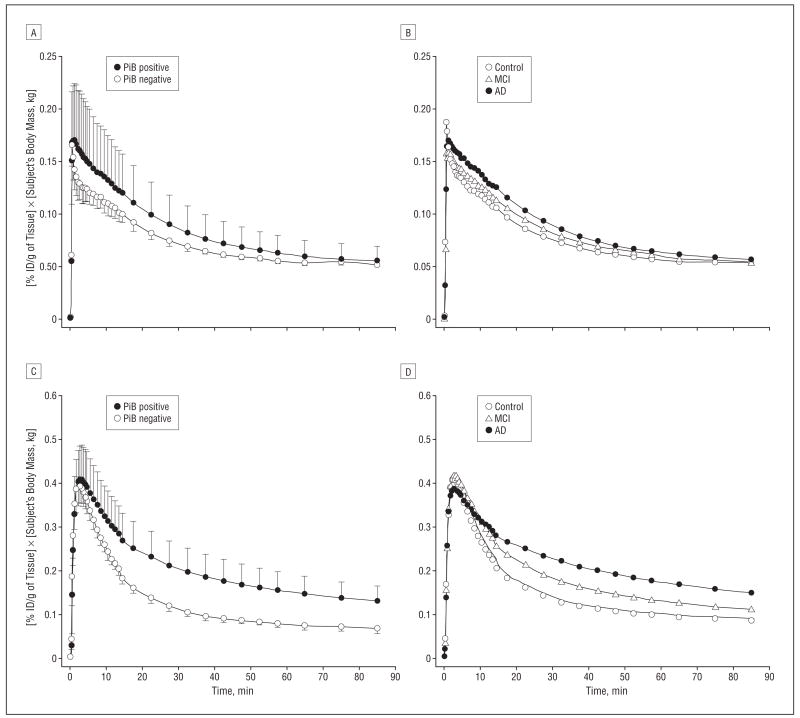
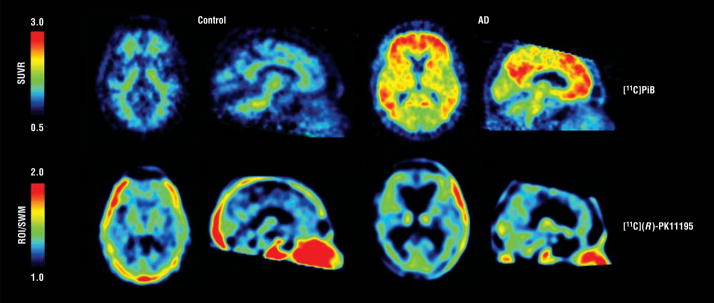
Results: Consistent with prior reports, subjects with a clinical diagnosis of probable AD showed significantly greater levels of [11C]PiB retention than control subjects, whereas patients with mild cognitive impairment spanned a range from control-like to AD-like levels of [11C]PiB retention. Additionally, 2 asymptomatic control subjects also exhibited evidence of elevated PiB retention in regions associated with the early emergence of plaques in AD and may represent prodromal cases of AD. We observed no differences in brain [11C](R)-PK11195 retention when subjects were grouped by clinical diagnosis or the presence or absence of beta-amyloid pathological findings as indicated by analyses of [11C]PiB retention.
Conclusions: These findings suggest that either microglial activation is limited to later stages of severe AD or [11C](R)-PK11195 is too insensitive to detect the level of microglial activation associated with mild to moderate AD.
Figures


References
-
- Mirra SS, Heyman A, McKeel D, et al. The Consortium to Establish a Registry for Alzheimer’s Disease (CERAD), part II: standardization of the neuropathologic assessment of Alzheimer’s disease. Neurology. 1991;41(4):479–486. - PubMed
-
- McGeer EG, McGeer PL. Inflammatory processes in Alzheimer’s disease. Prog Neuropsychopharmacol Biol Psychiatry. 2003;27(5):741–749. - PubMed
-
- Hyman BT, West HL, Rebeck GW, Lai F, Mann DM. Neuropathological changes in Down’s syndrome hippocampal formation: effect of age and apolipoprotein E genotype. Arch Neurol. 1995;52(4):373–378. - PubMed
-
- Mintun MA, Larossa GN, Sheline YI, et al. [11C]PIB in a nondemented population: potential antecedent marker of Alzheimer disease. Neurology. 2006;67(3):446–452. - PubMed
-
- Pike KE, Savage G, Villemagne VL, et al. Beta-amyloid imaging and memory in non-demented individuals: evidence for preclinical Alzheimer’s disease. Brain. 2007;130(pt 11):2837–2844. - PubMed
Publication types
MeSH terms
Substances
Grants and funding
- K24 MH001717/MH/NIMH NIH HHS/United States
- R01 MH071151/MH/NIMH NIH HHS/United States
- R01 MH064921/MH/NIMH NIH HHS/United States
- R01 AG018402/AG/NIA NIH HHS/United States
- AG025204/AG/NIA NIH HHS/United States
- R37 AG025516/AG/NIA NIH HHS/United States
- P50 AG005133/AG/NIA NIH HHS/United States
- RF1 AG025516/AG/NIA NIH HHS/United States
- P01 AG025204/AG/NIA NIH HHS/United States
- MH01717/MH/NIMH NIH HHS/United States
- R01 MH070729/MH/NIMH NIH HHS/United States
- S10 RR014772/RR/NCRR NIH HHS/United States
- AG05133/AG/NIA NIH HHS/United States
- AG025516/AG/NIA NIH HHS/United States
- R21 AG025829/AG/NIA NIH HHS/United States
- R01 AG025516/AG/NIA NIH HHS/United States
- AG025829/AG/NIA NIH HHS/United States
- AG018402/AG/NIA NIH HHS/United States
- MH070729/MH/NIMH NIH HHS/United States
LinkOut - more resources
Full Text Sources
Other Literature Sources
Medical
