. 2009 Feb;119(2):376-86.
doi: 10.1172/JCI36587. Epub 2009 Jan 12.

Loss of viral fitness and cross-recognition by CD8+ T cells limit HCV escape from a protective HLA-B27-restricted human immune response

Affiliations

Loss of viral fitness and cross-recognition by CD8+ T cells limit HCV escape from a protective HLA-B27-restricted human immune response

Eva Dazert et al. J Clin Invest. 2009 Feb.

Abstract

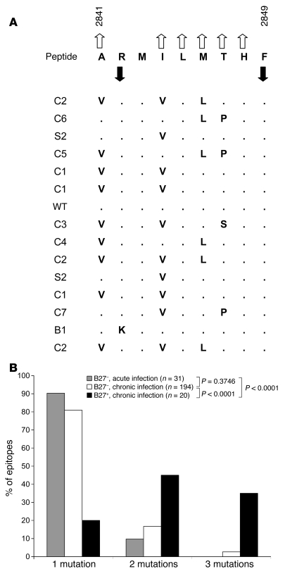
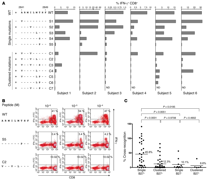
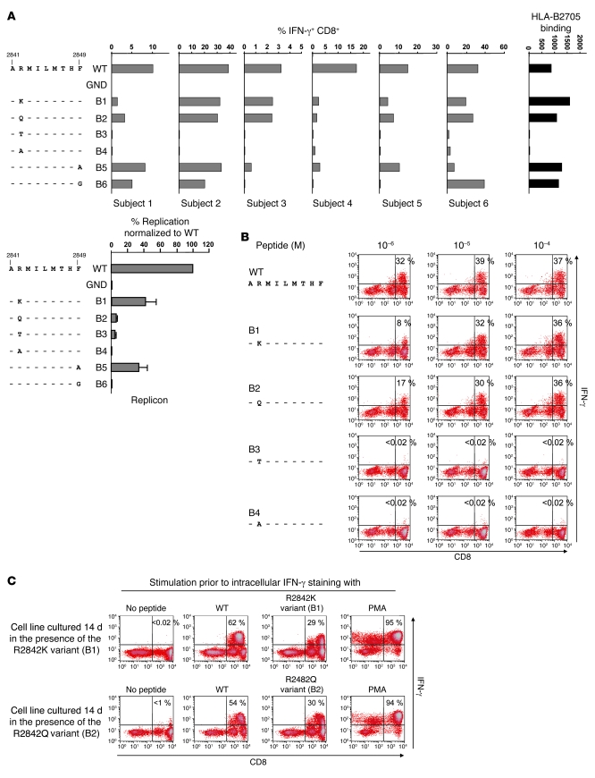
There is an association between expression of the MHC class I molecule HLA-B27 and protection following human infection with either HIV or HCV. In both cases, protection has been linked to HLA-B27 presentation of a single immunodominant viral peptide epitope to CD8+ T cells. If HIV mutates the HLA-B27-binding anchor of this epitope to escape the protective immune response, the result is a less-fit virus that requires additional compensatory clustered mutations. Here, we sought to determine whether the immunodominant HLA-B27-restricted HCV epitope was similarly constrained by analyzing the replication competence and immunogenicity of different escape mutants. Interestingly, in most HLA-B27-positive patients chronically infected with HCV, the escape mutations spared the HLA-B27-binding anchor. Instead, the escape mutations were clustered at other sites within the epitope and had only a modest impact on replication competence. Further analysis revealed that the cluster of mutations is required for efficient escape because a combination of mutations is needed to impair T cell recognition of the epitope. Artificially introduced mutations at the HLA-B27-binding anchors were found to be either completely cross-reactive or to lead to substantial loss of fitness. These results suggest that protection by HLA-B27 in HCV infection can be explained by the requirement to accumulate a cluster of mutations within the immunodominant epitope to escape T cell recognition.


Figures

Figure 1
Figure 1. Escape mutations within the NS5B2841–2849 epitope are clustered and spare HLA-B27–binding anchors.
(A) Amino acid sequence of NS5B2841–2849. Upward-pointing open arrows indicate residues that are predicted to interact with the TCR; downward-pointing filled arrows indicate HLA-B27–binding anchors (16). Autologous viral sequences found in 15 HLA-B27+ patients with chronic genotype 1 HCV infection are shown below; points indicate homologous residues (8). (B) Numbers of amino acid substitutions per escape variant are compared in HLA-B27+ and HLA-B27 subjects. Sequences for HLA-B27 subjects were taken from the literature: for acute HCV infection, longitudinal sequences or sequences from a known donor and the patient were compared (–27), while for chronic HCV infection, patient sequences were compared with genotype/subtype-matched consensus sequences (, –33). Sequences in HLA-B27+ patients with chronic HCV infection were included from this study (Figure 1A) and from a previous, independent report (32). P values were calculated using Mann-Whitney U test.
Figure 2
Figure 2. Clustering of mutations at TCR contact sites is required for efficient escape from the WT-specific T cell response.
(A) PBMCs from 6 HLA-B27+ individuals with HCV infection that resolved either spontaneously or through therapy were cultured for 14 days in the presence of WT peptide NS5B2841–2849. Cells were then tested for IFN-γ production after 5 hours stimulation with WT or variant peptides at a concentration of 10–5 M. Variants with single or clustered substitutions are grouped. A plus indicates that this mutation occurs in vivo, while a minus indicates that this mutation was not found in vivo. ND, not done. (B) Representative dot blots corresponding to Figure 2A (subject 2) are shown, including titration of the peptides in different concentrations as indicated. Percentages indicate IFN-γ+CD8+ T cells. (C) Levels of cross-recognition observed for each single and clustered escape variant of the HLA-B27–restricted NS5B2841–2849 epitope are compared with the levels of cross-recognition of a number of single or clustered escape variants in HLA-B27 patients described previously by our group (29). The level of cross-recognition for each variant was calculated by dividing the response to the variant peptide at a concentration of 10–5 M by the response to WT peptide at a concentration of 10–5 M. P values were calculated using Mann-Whitney U test. Bars denote mean.
Figure 4
Figure 4. Variants with substitutions at the HLA-B27–binding anchors are either cross-recognized or show limited replication competent.
(A) PBMCs from 6 HLA-B27+ individuals with HCV infection that resolved either spontaneously or through therapy were cultured for 14 days in the presence of WT peptide NS5B2841–2849. Cells were then tested for IFN-γ production after 5 hours stimulation with WT or variant peptides containing substitutions at HLA-B27–binding anchors P2 or P9 at a concentration of 10–5 M. The far-right panel shows HLA-B2705 binding of the different peptides, and the lower-left panl shows the replicon data for anchor residue mutants 48 hours after electroporation. WT was normalized to 100%. Data are presented as mean ± SD. (B) Representative dot plots corresponding to A (subject 2) are shown, including titration of the peptides in different concentrations as indicated. Percentages indicate IFN-γ+CD8+ T cells. (C) Cell lines cultured for 14 days in the presence of the R2842K (upper panels) or R2842Q (lower panels) variant peptides showed high amounts of IFN-γ–producing CD8+ T cells after stimulation with WT or the respective variant peptide for 5 hours. Percentages indicate IFN-γ+CD8+ T cells.
Figure 3
Figure 3. Clustered mutations at the TCR contact sites do not substantially impair replicative capacity.
(A) Schematic overview of the subgenomic HCV reporter replicon pFKi341PiLucNS3-3ιET. Luc, luciferase gene of the firefly Photinus vulgaris; P-I, polio virus internal ribosomal entry site; E-I, encephalomyocarditis virus internal ribosomal entry site. Asterisks indicate adaptive mutations (E1202G, T1280I, K1846T). Epitope NS5B2841–2849 is highlighted. Black arrows correspond to TCR residues, gray arrows to anchor residues. (B) Representative time course of mutants S2, C1, and C3 compared with those of WT and the negative mutant GND. Replicative capacity is expressed as percentage after normalization to the 4-hour input RNA value that reflected transfection efficiency. (C) Results for all mutants at 48 hours. WT was normalized to 100%. Variants with single or clustered substitutions are grouped. A plus indicates that this mutation occurs in vivo, while a minus indicates that this mutation was not found in vivo. Data are presented as mean ± SD.
Figure 5
Figure 5. Structural analysis of the epitope.
(A) NS5B displayed as ribbons and colored according to individual domains (red: fingers; yellow: palm; blue: thumb). The epitope is shown in blue-green. (B) Close-up of the epitope region. Residues mutated in this study are shown in stick representation and colored according to atom type (N: blue; O: red; S: orange; C: blue-green). Four of these residues are labeled. Also labeled are helices Q (which harbors the epitope) and A (from the “fingertips”), as well as non-nucleoside inhibitor (NNI) site B. Note that helix Q is both central to the thumb’s folding domain and involved in the NNI pockets’ buildup. (C) Interactions of R2842 with neighboring residues. (D and E) Models of the same region after replacement of R2842 with lysine (variant B1) or glutamine (variant B2). (F) Interactions of I2844 with neighboring residues. (G) Model of the variant S2 containing the I2844V substitution. (H) Interactions of T2847 with neighboring residues. In C, F, and H, the neighboring residues are shown as sticks, with carbons in white; hydrogen bonds are represented as dotted yellow lines. (I and J) Models of the variants containing substitution T2847S (S4) and T2847P (S5), respectively.

References

    1. Lauer G.M., Walker B.D. Hepatitis C virus infection. N. Engl. J. Med. 2001;345:41–52. doi: 10.1056/NEJM200107053450107. - DOI - PubMed
    1. Dustin L.B., Rice C.M. Flying under the radar: the immunobiology of hepatitis C. Annu. Rev. Immunol. 2007;25:71–99. doi: 10.1146/annurev.immunol.25.022106.141602. - DOI - PubMed
    1. Lechner F., et al. Analysis of successful immune responses in persons infected with hepatitis C virus. J. Exp. Med. 2000;191:1499–1512. doi: 10.1084/jem.191.9.1499. - DOI - PMC - PubMed
    1. Thimme R., et al. Determinants of viral clearance and persistence during acute hepatitis C virus infection. J. Exp. Med. 2001;194:1395–1406. doi: 10.1084/jem.194.10.1395. - DOI - PMC - PubMed
    1. Shoukry N.H., et al. Memory CD8+ T cells are required for protection from persistent hepatitis C virus infection. J. Exp. Med. 2003;197:1645–1655. doi: 10.1084/jem.20030239. - DOI - PMC - PubMed

Publication types