. 2009 Mar;89(3):301-14.
doi: 10.1038/labinvest.2008.168. Epub 2009 Jan 12.

PAX5 is expressed in small-cell lung cancer and positively regulates c-Met transcription

Affiliations

PAX5 is expressed in small-cell lung cancer and positively regulates c-Met transcription

Rajani Kanteti et al. Lab Invest. 2009 Mar.

Abstract

PAX5 is a nuclear transcription factor required for B cell development, and its expression was evaluated in upper aerodigestive malignancies and pancreatic cancer by immunoblotting. The PAX5 protein expression was relatively strong in small-cell lung cancer (SCLC, 11/12); however, its expression was not detected in non-SCLC (NSCLC, n=13), mesothelioma (n=7), pancreatic (n=6), esophageal (n=6) and head and neck cancer cell lines (n=12). In comparison, PAX8 and PAX3 expressions were absent or non-detectable in SCLC cell lines; however, PAX8 was expressed in most of the tested NSCLC cell lines (13/13) and also frequently in all the other cell lines. We also detected frequent expressions of PAX2 and PAX9 protein in the various cell lines. Utilizing neuroendocrine tumor samples, we found that the frequency as well as the average intensity of the expression of PAX5 increased from pulmonary carcinoid (9%, moderate and strong PAX5 expression, n=44), to large-cell neuroendocrine carcinoma (LCNC, 27%, n=11) to SCLC (33%, n=76). FISH analysis revealed no translocations of the PAX5 gene, but polyploidy in some SCLC tumor tissues (6/37). We determined that PAX5 could regulate the transcription of c-Met using luciferase-coupled reporter and chromatin immunoprecipitation analysis. In addition, the phospho-c-Met (active form) and PAX5 were both localized to the same intra-nuclear compartment in hepatocyte growth factor treated SCLC cells and interacted with each other. Finally, we determined the therapeutic translational potential of PAX5 using PAX5 knockdown SCLC cells in conjunction with Topoisomerase 1 (SN38) and c-Met (SU11274) inhibitors. Loss of endogenous PAX5 significantly decreased the viability of SCLC cells, especially when combined with SN38 or SU11274, and maximum effect was seen when both inhibitors were used. Therefore, we propose that PAX5 could be an important regulator of c-Met transcription and a potential target for therapy in SCLC.


Figures

Fig. 1
Fig. 1. Expression profile of PAX5 in SCLC, NSCLC, Mesothelioma, Esophagus, Pancreas, and Head and Neck cancers
Each panel represents a collection of cancer cell lines that signify a particular cancer. The whole cell lysates were immunoblotted with anti-PAX5, anti-PAX2, anti-PAX8 and anti-PAX9 antibodies. The same sample set was also used in parallel to determine c-Met, RON and Topo1 expression using specific antibodies in immunoblotting. β-actin level served as loading control.
Fig. 2
Fig. 2. Expression of PAX5 in lung tumor tissues of neuroendocrine origin
A. Nuclear PAX5 staining was carried out as described in Materials and Methods. Immunohistochemistry images represent negative (0), weak (1+), moderate (2+) and strongly positive (3+) PAX5 expression of different tumor subtypes. B–D. Pie-charts show relative proportion of negative (blue), weak positive (yellow), moderate positive (red) and strong positive cases (maroon) in carcinoid, LCNC and SCLC tumor samples.E. PAX5 expression levels and relative frequency based on independent scoring by two pathologists. It was lowest in lung carcinoids (top panel, 73% negative cases, n=44), intermediate in large cell neuroendocrine carcinomas (middle panel, 45% negative cases, n=11), and highest in small cell carcinomas (bottom panel, 34% negative cases, n=76). A chi-square test shows statistically significant increased PAX5 expression from carcinoid to LCNC to SCLC (P=0.007).
Fig. 3
Fig. 3. PAX5 locus on human chromosome 9 and FISH analysis
A. The PAX5 gene, located on chromosome 9 on p13, and involved in t(9; 14)(p13;q32) translocations recurring in small lymphocytic lymphomas of the plasmacytoid subtype. B. The PAX5 gene locus and the digitonin and biotin labeled BAC constructs used in the FISH are represented schematically. C. Flowchart shows SCLC tissue sample processing for FISH analysis. D. Representative micrograph of a SCLC tissue FISH analysis.
Fig. 4
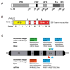
Fig. 4. PAX5 gene mutational analysis
A. PAX5 functional domains such as PD, DNA binding paired domain; OP, conserved octapeptide; HD, partial homeodomain; TA, transactivation domain; ID, inhibitory domain are schematically represented. B. Exons that code various domains in PAX5 protein are shown. C. Summary of mutations detected in SCLC cell lines and tissues. The PAX5 protein coding exons were PCR amplified and sequenced using genomic DNA from 21 SCLC cell lines and 24 SCLC tumor tissues. A non-synonymous heterozygous mutation 964G>AG resulting in an amino acid change A322S of the TA domain (amino acids 304–358) was detected in cell line H146. All the other mutations were synonymous.
Fig. 5
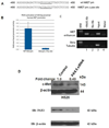
Fig. 5. PAX5 is a direct activator of c-Met transcription
A. PAX5 responsive element in the MET receptor gene. Sequence of the MET1 paired consensus site in the MET promoter and in the reporter construct wt hMET pm is shown. The sequence below has the MET1 site destroyed, GTCCCGC to ACTAGTC. B. Transfection of HEK 293T cells with both the hMET pm (wild type MET promoter sequence) and hMET pm D (MET promoter with MET1 site mutated) reporter constructs were carried out and all transfections were normalized for transfection efficiency and the reporter activities were expressed as fold activation compared with transfection without PAX5. (mean +/− SD, n=12 for each set). C. Chromatin Immunoprecipitation (ChIP) analysis was performed in H69 SCLC cells. The top panel represents PCR product obtained using primers for the MET promoter enhancer and the bottom panel represents PCR product using primers located in exon 4 of the β-tubulin gene (negative control). Lane 1, precipitation with mouse anti-PAX5 antibody; Lane 2, precipitation with normal mouse IgG; Lane 3, cell lysate input, pre-ChIP; and Lane 4, water blank PCR negative control are shown. D. H526 SCLC were transfected with scrambled and PAX5 specific siRNA (four different siRNAs pooled) at 100 nM. As shown, loss of PAX5 protein expression in siRNA treated cells is accompanied by a similar loss in c-Met protein levels.
Fig. 6
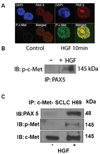
Fig. 6. PAX5 interacts with activated c-Met in SCLC cells upon HGF treatment
A. Representative confocal pictures demonstrating individual staining patterns for phospho-c-Met and PAX5 and the two merged in untreated (left quadrant) and HGF treated (right quadrant) in H82 cells are shown. The cells were treated for 10 min with HGF (40ng/ml). B. Co-immunoprecipitation of phospho-c-Met with PAX5. H249 cells were treated with and without HGF (40ng/ml) for 10 minutes and cell lysates prepared by standard method. Immunoprecipitation ((200µg lysates) was carried out using anti-PAX5 antibody that revealed significant amounts of phosphor-c-Met only in HGF treated sample. C. Immunoprecipitation of PAX5 coprecipitated significant amounts of PAX5.
Fig. 7
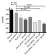
Fig. 7. Combinatorial effect of PAX5 knock down with cMet and Topo1 inhibitors in SCLC cells
H69 SCLC cells were transfected with either control or PAX5 specific siRNA duplexes (Qiagen). As shown, they were then treated with either SU11274, or SN38, or a combination of both. The histograms represent percentage of cell viability along with statistical significance.

References

    1. Abidoye O, Ferguson MK, Salgia R. Lung carcinoma in African Americans. Nat Clin Pract Oncol. 2007;4(4):118–129. - PubMed
    1. Robson EJD, He S-J, Eccles MR. A panorama of PAX genes in cancer and development. Nature Reviews Cancer. 2006;6:52–62. - PubMed
    1. Cobaleda C, Schebesta A, Delogu A, et al. Pax5: the guardian of B cell identity and function. Nat Immunol. 2007;8:463–470. - PubMed
    1. Krenacs L, Himmelmann AW, Quintanilla-Martinez L, et al. Transcription factor B-cell-specific activator protein (BSAP) is differentially expressed in B cells and in subsets of B-cell lymphomas. Blood. 1998;A92:1308–1316. - PubMed
    1. Kozmik Z, Sure U, Rüedi D, et al. Deregulated expression of PAX5 in medulloblastoma. Proc Natl Acad Sci U S A. 1995;92:5709–5713. - PMC - PubMed

Publication types

MeSH terms

Substances