. 2009 May-Jun;87(4):324-36.
doi: 10.1038/icb.2008.103. Epub 2009 Jan 13.

Clustering T-cell GM1 lipid rafts increases cellular resistance to shear on fibronectin through changes in integrin affinity and cytoskeletal dynamics

Affiliations

Clustering T-cell GM1 lipid rafts increases cellular resistance to shear on fibronectin through changes in integrin affinity and cytoskeletal dynamics

Jason S Mitchell et al. Immunol Cell Biol. 2009 May-Jun.

Abstract

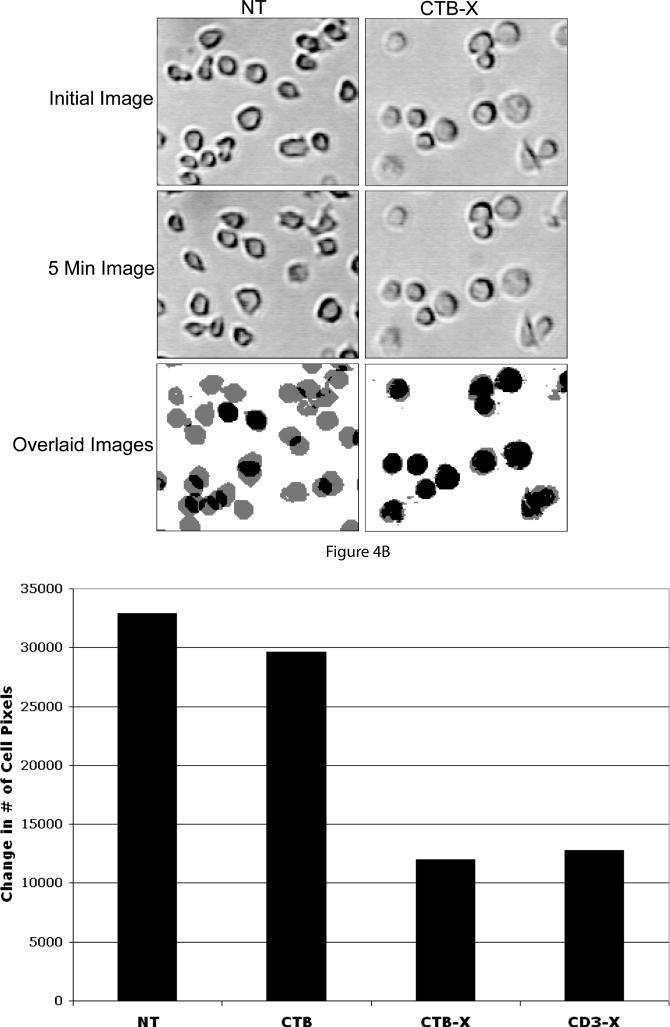
Lipid rafts are small laterally mobile microdomains that are highly enriched in lymphocyte signaling molecules. GM1 gangliosides are a common lipid raft component and have been shown to be important in many T-cell functions. The aggregation of specific GM1 lipid rafts can control many T-cell activation events, including their novel association with T-cell integrins. We found that clustering GM1 lipid rafts can regulate beta1 integrin function. This was apparent through increased resistance to shear flow-dependent detachment of T cells adherent to the alpha4beta1 and alpha5beta1 integrin ligand fibronectin (FN). Adhesion strengthening as a result of clustering GM1 enriched lipid rafts correlated with increased cellular rigidity and morphology through the localization of cortical F-actin, the resistance to shear-induced cell stretching, and an increase in the surface area and symmetry of the contact area between the cell surface and adhesive substrate. Furthermore, clustering GM1 lipid rafts could initiate integrin 'inside-out' signaling mechanisms. This was seen through increased integrin-cytoskeleton associations and enhanced soluble binding of FN and VCAM-1, suggesting the induction of high-affinity integrin conformations. The activation of these adhesion-strengthening characteristics appears to be specific for the aggregation of GM1 lipid rafts as the aggregation of the heterogeneous raft-associated molecule CD59 failed to activate these functions. These findings indicate a novel mechanism to signal to beta1 integrins and to activate adhesion-strengthening processes.


Figures

Figure 1
Figure 1. Clustering GM1 Lipid Rafts Increases T cell Resistance to Shear Flow Detachment and Stretching
A) Jurkat T cells were treated as indicated for 5 mins, perfused into a parallel plate flow chamber assembled with FN (5 ug/ml) adsorbed slides, and allowed to settle for 10 min. A linear gradient of shear flow was applied to the adhered cells and the % cells remaining were enumerated every 20 sec. The data presented is the percentage mean cells remaining from triplicate runs +/− SD. B) Images were taken at the indicated shear values from two runs from A). C) Images from the runs were taken at the indicated shear values from A) and the stretch index for all the cells in each image was determined and averaged as described in the materials and methods.
Figure 2
Figure 2. Clustering GM1 Lipid Rafts Induces Cortical F-actin Polymerization
Jurkat T cells were treated with the indicated stimuli, plated onto FN (5ug/ml) adsorbed glass coverslips, and allowed to adhere for 20 min. Cells were fixed, permeabilized, and stained for F-actin polymerization with Alexa Fluor 594 labeled phalloidin. Arrows indicate regions of F-actin polarization in migrating T cells. The images presented are representative from three separate experiments.
Figure 3
Figure 3. Clustering GM1 Lipid Rafts Increases Surface Area and Symmetry of Contact Area
Jurkat T cells were treated as indicated, plated onto FN (5ug/ml) adsorbed glass coverslips, and allowed to adhere for 20 min. The cells were fixed and images at the contact plane were obtained with a confocal microscope using IRM technology. Black areas are considered strong focal contacts (black arrow) gray areas close contacts (white arrow), and white areas are non-contacts. The images presented are representative pictures from three separate experiments.
Figure 4
Figure 4. Clustering GM1 Lipid Rafts Inhibits T Cell Mobility
A) Jurkat T cells were treated as indicated, and added to FN coated wells for 20 min. After incubation, images of the cells were taken at the initial timepoint and then again after 5 min had elapsed. Mobile cells are shown as gray images in the overlaid image, while non-mobile cells are shown in black. B) Cellular mobility is quantified by counting the number of cellular pixels in the initial and overlaid images, and is represented as the change in cellular pixels from the overlaid and the initial images. Results are representative of two experiments performed.
Figure 5
Figure 5. CD3 and GM1 Lipid Raft Clustering Inhibits Chemotaxis through p56lck
A) Jurkat or B) JCAM 1.6 and JCAM 1.6 reconstituted with p56lck T cells were stimulated with antibody complexes as indicated and placed in FN coated transwells (10 ug/ml) with or without 15 ng/ml SDF-1α in the bottom well. The degree of cellular migration was determined by counting the number of cells in the bottom wells.
Figure 6
Figure 6. Clustering GM1 Lipid Rafts Increases T cell Integrin Affinity
A) Jurkat T cells were pretreated with 125I labeled FN for 10 min and then treated as indicated for 30 min. Triplicate cell pellets were counted in a gamma counter to determine relative sFN binding. The data is shown as the mean CPM +/− SD from one experiment. (*, p < 0.001 using paired t test to compare rCTB to rCTB-X from three independent experiments. B) Jurkat T cells were pretreated with inhibitors RGD (20ug/ml) and 772 (100uM) and then used in the sFN binding assay as described in A). Results are representative of two experiments performed. C) Jurkat T cells were pretreated for 10 min with recombinant sVCAM-1 (5ug/ml) and then treated as indicated. At different timepoints aliquots were rapidly diluted, immediately fixed, and sVCAM-1 binding was detected by flow cytometry. The experiments were done in triplicate and the data is presented as the mean increase in MFI from the inital time point before the stimulus was added, +/− SD.
Figure 7
Figure 7. Clustering GM1 Lipid Rafts Increases β1 Integrin Anchorage to the Cytoskeleton
A) Crosslinked β1 integrins acquire resistance to NP-40 solubilization. Jurkat T cells were incubated with Alexa Fluor 488 labeled anti-β1 integrin mAb 33B6 on ice for 30 min, and either uncrosslinked (33B6) or crosslinked with an isotype specific antibody (33B6-X). Cells were then untreated or were briefly treated with 0.05% NP-40 to solubilize and wash way unanchored integrin. The flow histograms are representative of three independent experiments. B) The % NP-40 resistant β1 integrin from A) is determined by dividing the MFI of NP-40 treated cells by the MFI of the cells that did not receive NP-40 treatment. C) Jurkat T cells were incubated as indicated at 37°C for 20 min and then recieved +/− NP-40 treatment to determine the % NP-40 resistant β1 integrin. The data shown is from three independent experiments and is presented as the mean increase in % NP-40 treatment from NT +/− SD (*, p < 0.001, ANOVA).
Figure 8
Figure 8. Illustration of the Effect of Contact Symmetry on Shear Resistance
IRM images show that unstimulated T cells produce non-symmetrical areas of contact while GM1 lipid raft clustered cells produce circular and symmetric regions of contact. In unstimulated cells, the offset between the center of resistance (ResistiveForceaxis) and the center of applied force (ShearForceaxis) will impose torsional moments on the cells. In cells where the contact area is symmetrical, the center of resistance and center of applied force are inline with each other and no torsional moments are created.

References

    1. Grabovsky V, Feigelson S, Chen C, Bleijs DA, Peled A, Cinamon G, et al. Subsecond induction of alpha4 integrin clustering by immobilized chemokines stimulates leukocyte tethering and rolling on endothelial vascular cell adhesion molecule 1 under flow conditions. J Exp Med. 2000;192:495–506. - PMC - PubMed
    1. Feigelson SW, Grabovsky V, Winter E, Chen LL, Pepinsky RB, Yednock T, et al. The Src kinase p56(lck) up-regulates VLA-4 integrin affinity. Implications for rapid spontaneous and chemokine-triggered T cell adhesion to VCAM-1 and fibronectin. J Biol Chem. 2001;276:13891–901. - PubMed
    1. Laudanna C, Alon R. Right on the spot. Chemokine triggering of integrin-mediated arrest of rolling leukocytes. Thromb Haemost. 2006;95:5–11. - PubMed
    1. Simons K, Ikonen E. Functional rafts in cell membranes. Nature. 1997;387:569–72. - PubMed
    1. Foger N, Marhaba R, Zoller M. Involvement of CD44 in cytoskeleton rearrangement and raft reorganization in T cells. J Cell Sci. 2001;114:1169–78. - PubMed

Publication types