. 2008 Dec 18;456(7224):976-979.
doi: 10.1038/nature07422.

A structural explanation for the binding of endocytic dileucine motifs by the AP2 complex

Affiliations

A structural explanation for the binding of endocytic dileucine motifs by the AP2 complex

Bernard T Kelly et al. Nature. .

Abstract

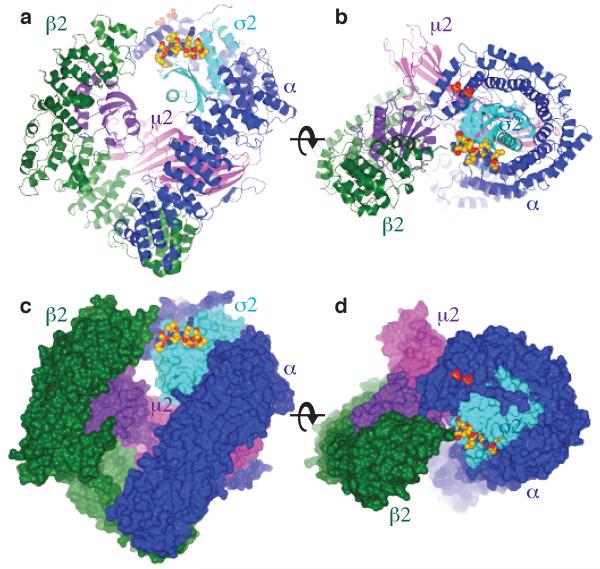
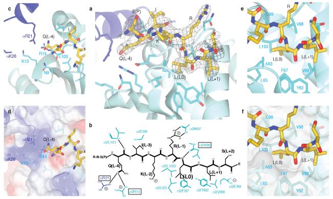
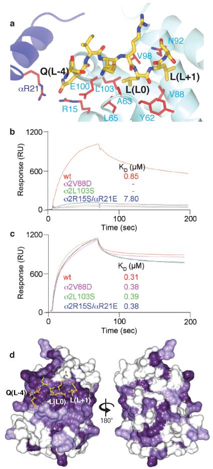
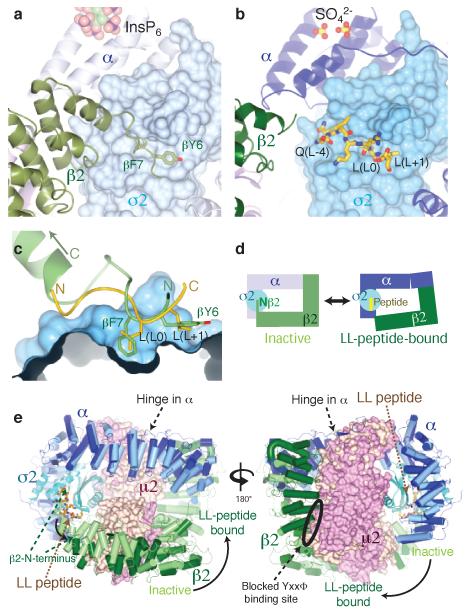
Most transmembrane proteins are selected as transport vesicle cargo through the recognition of short, linear amino acid motifs in their cytoplasmic portions by vesicle coat proteins. In the case of clathrin-coated vesicles (CCVs) the motifs are recognised by clathrin adaptors. The AP2 adaptor complex (subunits α,β2,μ2,σ2) recognises both major endocytic motifs: YxxΦ motifs and [DE]xxxL[LI] acidic dileucine motifs. Here we describe the binding of AP2 to the endocytic dileucine motif from CD4 . The major recognition events are the two leucine residues binding in hydrophobic pockets on σ2. The hydrophilic residue four residues upstream from the first leucine sits on a positively charged patch made from residues on σ2 and α subunits. Mutations in key residues inhibit the binding of AP2 to ‘acidic dileucine’ motifs displayed in liposomes containing PtdIns4,5P2, but do not affect binding to YxxΦ motifs via μ2. In the ‘inactive’ AP2 core structure , both motif binding sites are blocked by different parts of the β2 subunit. To allow a dileucine motif to bind, the β2 N-terminus is displaced and becomes disordered; however, in this structure the YxxΦ binding site on μ2 remains blocked.


Figures

Figure 1
Figure 1. Structure of the AP2 adaptor core in complex with the dileucine peptide from CD4
Orthogonal views of the dileucine motif liganded AP2 complex in ribbon (a and b) and in molecular surface (c and d) representations. In a and c the membrane is parallel to the upper face of the complex and in b and d the complex is viewed through the membrane with the membrane interacting surface facing up. The α subunit is coloured dark blue, β2 green, σ2 pale blue, N-μ2 purple and C-μ2 mauve. The dileucine motif peptide, shown as spheres with carbons coloured gold. The two sulphate groups bound to the α subunit in the PtdInsP2 site are shown.
Figure 2
Figure 2. Details of binding of the CD4 dileucine signal by the σ2 and α subunits of AP2
a The dileucine peptide is bound mainly by the pale blue σ2 subunit near its interface with the α subunit (dark blue). The peptide is shown in its final 2mFo-dFc electron density (cropped around the peptide and contoured at 0.11 e/Å3). b Schematic representation of the dileucine peptide with the principal side chains involved in its binding. Mutants in the boxed residues were kinetically analysed. c and d details of the polar Q(L-4) binding pocket: d with semi transparent electrostatic surface representation (coloured from red −0.5V to blue +0.5V), showing the positive charge principally due to αR21, σ2K13 and σ2R15. e and f: the deep and shallow pockets on σ2 involved in recognising the leucine residues at position L0 and L+1. Labelled side chains are in the σ2 subunit unless otherwise indicated.
Figure 3
Figure 3. Confirmation of location and conservation amongst different σ subunits of the dileucine motif binding site
a The dileucine peptide binding site with residues whose mutation strongly inhibit dileucine peptide binding whilst not affecting YxxΦ motif binding are coloured red (See Supplementary Table S1 and Figure S8). b and c Sensorgrams and KD values for binding of wild type AP2 core and three mutants thereof that strongly inhibit binding of AP2 to PtdIns4,5P2 containing liposomes displaying the CD4 Q peptide motif (b) but do not effect binding to PtdIns4,5P2 containing liposomes displaying the TGN38 YxxΦ motif (c). d Mammalian σ2, σ1a, σ3 and σ4 were aligned using ClustalW (Figure S7) and the residue conservation plotted from dark purple (absolute conservation) to white (no conservation) onto the surface of mammalian σ2 in two views related by a rotation of 180°. The binding site for the dileucine motif peptide is the outstanding feature of surface residue conservation.
Figure 4
Figure 4. A conformational change in AP2 is required for dileucine peptide binding
In the IP6-liganded inactive conformation (a) the N-terminus of β2 is held in place by β2F7 and β2Y6 sitting in the L0 and L+1 pockets. For dileucine motifs to bind the N-terminus of β2 is displaced from the surface of σ2 (b). (c) close-up view of the dileucine binding site: note that the LL peptide (gold) runs in the opposite direction to the N-terminus of β2 (green). (d) Schematic representation of the conformational change: a 20° hinge movement in α moves the N-terminus of β2 out of the LL-binding site, allowing the motif to bind. The μ2 subunit has been omitted for clarity. (e) front & back views of the conformational change: the IP6-liganded inactive conformation is shown in pale colours, the unlocked dileucine-peptide bound conformation in dark colours. The YxxΦ site on μ2 remains blocked, and is remote from the dileucine binding site.

References

    1. Owen DJ, Evans PR. A structural explanation for the recognition of tyrosine-based endocytotic signals. Science. 1998;282:1327–32. - PMC - PubMed
    1. Pitcher C, Honing S, Fingerhut A, Bowers K, Marsh M. Cluster of differentiation antigen 4 (CD4) endocytosis and adaptor complex binding require activation of the CD4 endocytosis signal by serine phosphorylation. Mol Biol Cell. 1999;10:677–91. - PMC - PubMed
    1. Collins BM, McCoy AJ, Kent HM, Evans PR, Owen DJ. Molecular architecture and functional model of the endocytic AP2 complex. Cell. 2002;109:523–35. - PubMed
    1. Hurley JH, Lee S, Prag G. Ubiquitin-binding domains. Biochem J. 2006;399:361–72. - PMC - PubMed
    1. Pryor PR, et al. Molecular basis for the sorting of the SNARE VAMP7 into endocytic clathrin-coated vesicles by the ArfGAP Hrb. Cell. 2008 In Press. - PMC - PubMed

Additional References for online Materials and Methods

    1. Leslie AG. The integration of macromolecular diffraction data. Acta Crystallogr D Biol Crystallogr. 2006;62:48–57. - PubMed
    1. Evans P. Scaling and assessment of data quality. Acta Crystallogr D Biol Crystallogr. 2006;62:72–82. - PubMed
    1. Adams PD, et al. PHENIX: building new software for automated crystallographic structure determination. Acta Crystallogr D Biol Crystallogr. 2002;58:1948–54. - PubMed
    1. Emsley P, Cowtan K. Coot: Model-Building Tools for Molecular Graphics. Acta Crystallographica Section D - Biological Crystallography. 2004;60:2126–2132. - PubMed
    1. Painter J, Merritt EA. Optimal description of a protein structure in terms of multiple groups undergoing TLS motion. Acta Crystallogr D Biol Crystallogr. 2006;62:439–50. - PubMed

MeSH terms