. 2009 Mar;108(5):1226-36.
doi: 10.1111/j.1471-4159.2009.05871.x. Epub 2009 Jan 29.

Aminoglycoside-induced histone deacetylation and hair cell death in the mouse cochlea

Affiliations

Aminoglycoside-induced histone deacetylation and hair cell death in the mouse cochlea

Fu-Quan Chen et al. J Neurochem. 2009 Mar.

Abstract

Post-translational modification of histones is an important form of chromatin regulation impacting transcriptional activation. Histone acetyltransferases, for example, acetylate lysine residues on histone tails thereby enhancing gene transcription, while histone deacetylases (HDACs) remove those acetyl groups and repress gene transcription. Deficient histone acetylation is associated with pathologies, and histone deacetylase inhibitors have been studied in the treatment of cancer and neurodegenerative diseases. Here we explore histone acetylation in cochlear sensory cells following a challenge with gentamicin, an aminoglycoside antibiotic known to cause loss of auditory hair cells and hearing. The addition of the drug to organotypic cultures of the mouse organ of Corti decreased the acetylation of histone core proteins (H2A Ack5, H2B Ack12, H3 Ack9, and H4 Ack8) followed by a loss of sensory cells. Protein levels of HDAC1, HDAC3 and HDAC4 were increased while the histone acetyltransferases such as CREB-binding protein and p300 remained unchanged. We next hypothesized that protecting histone acetylation should prevent cell death and tested the effects of HDAC-inhibitors on the actions of gentamicin. Co-treatment with trichostatin A maintained near-normal levels of acetylation of histone core proteins in cochlear hair cells and attenuated gentamicin-induced cell death. The addition of sodium butyrate also rescued hair cells from damage by gentamicin. The results are consistent with an involvement of deficient histone acetylation in aminoglycoside-induced hair cell death and point to the potential value of HDAC-inhibitors in protection from the side effects of these drugs.


Conflict of interest statement

Disclosure Statement

There are no conflicts of interest for any of the authors.

The data contained in the manuscript have not been previously published, have not been submitted elsewhere, and will not be submitted elsewhere while under review.

All experimental protocols were approved by the University of Michigan Committee on Use and Care of Animals and animal care was supervised by the University of Michigan’s Unit for Laboratory Animal Medicine.

All authors have reviewed the contents of the manuscript, approve of its contents, and validate the accuracy of the data.

Figures
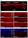
Figure 1
Figure 1. Gentamicin-induced hair cell loss in mouse organotypic cultures of the organ of Corti
A: A whole mount of the organ of Corti explanted at postnatal day 3 and maintained in culture for 2 days was stained for F-actin with rhodamine phalloidin (red fluorescence). The outer stained perimeter contains the hair cell layer. Basal, middle and apical segments indicate the approximate areas analyzed in this study. Scale bar = 500 μm. B: Base-to-apex gradient of hair cell loss. Surface preparations of the basal segment show the outlines of inner and outer hair cells stained for F-actin with rhodamine phalloidin. Control incubations in the absence of gentamicin maintained a normal appearance at 24 h. In the basal segment of the organ of Corti, outer hair cell loss began after 16 h of incubation with gentamicin and progressed with increasing time while outer hair cells at the apex (GM 20h Apex) remained intact. The images are representative of 8 individual preparations at each time point. Scale bar = 20 μm. C: Hensen’s cells remained intact after treatment with 0.2 mM gentamicin for 20h. Nuclei were stained blue with Hoechst 33342 and Hensen’s cells can be identified by their position on the surface preparation and in comparison with F-actin staining for hair cells. Abbreviations: GM, gentamicin; IHC, inner hair cells; OHC1, OHC2, OHC3, outer hair cells of the first, second and third row, respectively.
Figure 2
Figure 2. Dose-dependence of outer hair cell loss
Surface preparations of the entire length of the explant were evaluated quantitatively for hair cell loss as described in ‘Methods’. After 24 h of incubation the percentage of missing outer hair cells increases with increasing doses of gentamicin. Percent of hair cells missing at gentamicin concentrations > 0.05 mM is significantly different (P < 0.05) from controls. Controls, n = 7; 0.05 mM, n = 3; 0.075 mM, n = 5; 0.1 mM, n = 15; 0.2 mM, n = 9. Data presented as average ± SD.
Figure 3 A-D
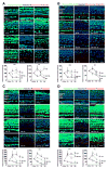
Figure 3 A-D. Acetylation of histone core proteins in both outer and inner hair cells is decreased by gentamicin and restored by trichostatin-A
Explants received the treatments as stated below and were stained (green) by immunocytochemistry for histone H3 Ack9 (A); histone H2A Ack5 (B); histone H2B Ack12 (C); or histone H4 Ack8 (D). Blue is Hoechst 33342 staining of nuclei. The figures are representative images of three individual preparations of the organ of Corti basal turn for each condition. Abbreviations: GM, gentamicin; vehicle, 0.2% DMSO; TSA, 200 nM trichostatin-A with 0.2% DMSO. IHC, inner hair cells; OHC1, outer hair cell first row; OHC2, outer hair cell second row; OHC3, outer hair cell third row. Scale bars = 20 μm. Left panels A, B, C, D: Explants were treated with 200 nM trichostatin-A for the times indicated on the images. ‘Vehicle’ denotes control incubation with 0.2% DMSO for 8 hr. Acetylation of histone is increased in the presence of trichostatin-A compared to the vehicle only. Middle panels A, B, C, D: Explants were treated with 0.2 mM gentamicin for the times indicated on the images. Acetylation of all histones in both outer and inner hair cells decreased in a time-dependent manner as compared to an 8-hr incubation in control medium. Right panels A, B, C, D: Explants received both 0.2 mM gentamicin and 200 nM trichostatin-A for the times indicated on the images. Histone acetylation in both outer and inner hair cells is maintained at near control levels for the first 8 hr (16 hr in the case of H3) of incubation but decreases at later times. Relative fluorescence intensity in both outer and inner hair cells obtained from the immunocytochemical images as described in the method section are stated at the bottom of each fluorescence image. Histones show similar patterns but different magnitude of decreased acetylation with gentamicin and increase with TSA at 8 h. Significance of differences: *: P<0.05 vs. Control; **: p<0.01 vs. Control.
Figure 4
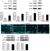
Figure 4. Histone deacetylases are transiently up-regulated by gentamicin
4A: Western blots detected histone deacetylases 1, -3, -4 and -6 in homogenates of organ of Corti explants. Membranes were sequentially stained for HDAC1 and HDAC3 and for HDAC4 and HDAC6. Significance of differences: *: p<0.05, **: p<0.01 compared to the corresponding control groups (n = 3, average ± SD). 4B: Immunocytochemistry of organ of Corti explants for histone deacetylase 1 (green) indicated a transient increase in outer hair cells at 8 h of gentamicin treatment. Blue is Hoechst 33342 staining for nuclei. The figure shows representative images from the basal turn of the organ of Corti (n = 3 at each time point). Scale bar = 20 μm. Abbreviations: GM, gentamicin; IHC, inner hair cell; OHC, outer hair cells; OHC1, outer hair cell first row; OHC2, outer hair cell second row; OHC3, outer hair cell third row; Hensen’s, hensen’s cell. 4C: Western blots detected histone acetylase CBP/p300 in homogenates of organ of Corti explants. Semi-quantitative analysis showed no difference between control and gentamicin treatment (n = 3, average ± SD).
Figure 5
Figure 5. Histone deacetylase inhibitors protect against gentamicin-induced hair cell loss
5A: Representative images from the basal turn of the organ of Corti after 20 h of incubation, showing the outline of hair cells stained with Alexa 488 phalloidin. Control: incubation with 0.2% DMSO; TSA: 200 nM trichostatin-A dissolved in 0.2% DMSO; GM 20h: explant treated with 0.2 mM gentamicin for 20 h; GM + vehicle: 0.2 mM gentamicin plus 0.2% DMSO; GM + TSA: 0.2 mM gentamicin plus 200 nM trichostatin-A dissolved in 0.2% DMSO. N = 8 for each condition. Scale bar = 20 μm. 5B: Quantitative evaluation of loss of outer and inner hair cells along the entire length of the organ of Corti explant after 20 h of incubation with gentamicin in the absence or presence of 200 nM trichostatin-A or 6 mM sodium butyrate. There is no inner or outer hair cell loss in control incubations with sodium sulfate (0.5 mM) substituting for gentamicin sulfate or with DMSO alone. N = 8 for each condition. Abbreviations: GM, gentamicin; SB, sodium butyrate; TSA, trichostatin-A. 5C: Average of hair cell survival between groups of Gentamicin, gentamicin plus SB, gentamicin plus DMSO, and gentamicin plus both DMSO and TSA (n = 8, average ± SD, *p<0.05, **p<0.01).
Figure 6
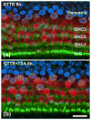
Figure 6. Trichostatin-A does not affect gentamicin uptake into outer hair cells
Explants were incubated for 8 h with 1.56 μg/mL gentamicin tagged with Texas Red (GTTR) in the absence (a) or presence (b) of 200 nM trichostatin-A. Red fluorescence is GTTR, green fluorescence is staining for F-actin in stereocilia of outer hair cells with Alexa 488-phalloidin, blue is Hoechst 33342 for nuclei. Images show a basal segment and are representative of three incubations under each condition. Scale bar = 20 μm.

References

    1. Bhaumik SR, Smith E, Shilatifard A. Covalent modifications of histones during development and disease pathogenesis. Nat Struct Mol Biol. 2007;14:1008–1016. - PubMed
    1. Dashwood RH, Ho E. Dietary histone deacetylase inhibitors: from cells to mice to man. Semin Cancer Biol. 2007;17:363–369. - PMC - PubMed
    1. de Ruijter AJ, van Gennip AH, Caron HN, Kemp S, van Kuilenburg AB. Histone deacetylases (HDACs): characterization of the classical HDAC family. Biochem J. 2003;370:737–749. - PMC - PubMed
    1. Ding D, Stracher A, Salvi RJ. Leupeptin protects cochlear and vestibular hair cells from gentamicin ototoxicity. Hear Res. 2002;164:115–126. - PubMed
    1. Drottar M, Liberman MC, Ratan RR, Roberson DW. The histone deacetylase inhibitor sodium butyrate protects against cisplatin-induced hearing loss in guinea pigs. Laryngoscope. 2006;116:292–296. - PMC - PubMed

Publication types

MeSH terms